Skip to Content
MilliporeSigma
All Photos(3)

Key Documents

C4606

Sigma-Aldrich

Anti-Calreticulin antibody produced in rabbit

IgG fraction of antiserum, buffered aqueous solution

Sign Into View Organizational & Contract Pricing


About This Item

MDL number:
UNSPSC Code:
12352203
NACRES:
NA.43

biological source

rabbit

Quality Level

conjugate

unconjugated

antibody form

IgG fraction of antiserum

antibody product type

primary antibodies

clone

polyclonal

form

buffered aqueous solution

mol wt

antigen 50 kDa

species reactivity

human, canine

technique(s)

immunoprecipitation (IP): suitable using whole cell RIPA lysate of the human epitheloid carcinoma HeLa cell line
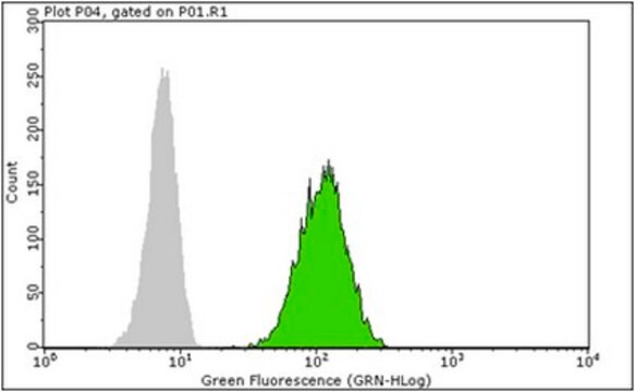
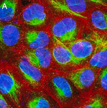
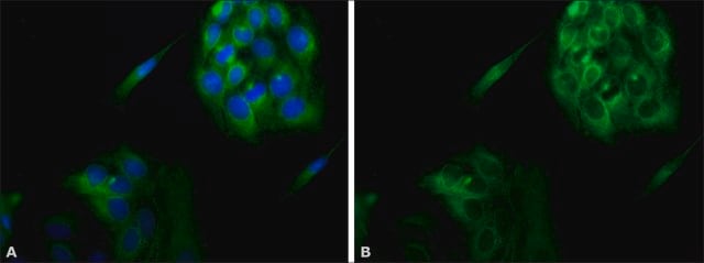
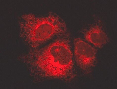
indirect immunofluorescence: 1:100 using 3% paraformaldehyde-fixed, 0.5% Triton X-100 treated, Madin Darby canine kidney MDCK cell line
microarray: suitable
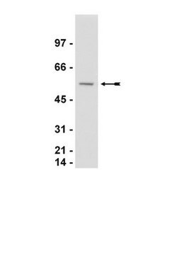
western blot: 1:2,000 using whole cell RIPA lysate of the human epitheloid carcinoma HeLa cell line

UniProt accession no.

shipped in

dry ice

storage temp.

−20°C

target post-translational modification

unmodified

Gene Information

human ... CALR(811)

General description

The gene for calreticulin (CALR) is located on the human chromosome 19p13.13. The encoded protein is a predominantly conserved calcium-binding chaperone, which is localized primarily to the lumen of the endoplasmic reticulum (ER). Calreticulin comprises three domains, a globular N-domain, a proline-rich P-domain, and a highly acidic C-domain.

Specificity

Anti-Calreticulin recognizes human and dog calreticulin (55-60 kDa).

Immunogen

synthetic peptide corresponding to the C-terminus of human calreticulin (amino acids 401-417).

Application

Anti-Calreticulin antibody produced in rabbit has been used in:
  • immunoblotting
  • immunofluorescence
  • immunocytochemistry{91
  • immunoprecipitation

Biochem/physiol Actions

Calreticulin facilitates transient interaction with newly synthesized cellular and extracellular proteins for folding and assembling in the endoplasmic reticulum (ER) before its localization to the cytosol or cell surface. Calreticulin acts as a lectin-like chaperone binding oligosaccharide residues of newly synthesized N-linked glycoproteins and misfolded proteins. It is believed to play a critical role in quality control processes during protein synthesis and folding and calcium (Ca2+) homeostasis. Increased expression of calreticulin increases the Ca2+ storage capacity of the ER. It also appears to modulate store-operated Ca2+-influx and to alter Ca2+ transport by the sarcoplasmic/ER Ca2+-adenosine triphosphatase (ATPase) (SERCA). Overexpression of calreticulin results in increased sensitivity of HeLa cells to drug-induced apoptosis. However, increased resistance to apoptosis has been observed in calreticulin-deficient cells.

Physical form

Solution in 0.01 M phosphate buffered saline, pH 7.4, containing 15 mM sodium azide.

Storage and Stability

Store at -20 °C. For continuous use, the product maybe stored at 2-8 °C for up to one month. For prolonged storage, freeze in working aliquots at-20 °C. Repeated freezing and thawing, or storage in “frost-free” freezers, is not recommended. If slight turbidity occurs upon prolonged storage, clarify the solution by centrifugation before use. Working dilution samples should be discarded ifnot used within 12 hours.

Disclaimer

Unless otherwise stated in our catalog or other company documentation accompanying the product(s), our products are intended for research use only and are not to be used for any other purpose, which includes but is not limited to, unauthorized commercial uses, in vitro diagnostic uses, ex vivo or in vivo therapeutic uses or any type of consumption or application to humans or animals.

Not finding the right product?  

Try our Product Selector Tool.

Storage Class

10 - Combustible liquids

wgk_germany

nwg

flash_point_f

Not applicable

flash_point_c

Not applicable


Certificates of Analysis (COA)

Search for Certificates of Analysis (COA) by entering the products Lot/Batch Number. Lot and Batch Numbers can be found on a product’s label following the words ‘Lot’ or ‘Batch’.

Already Own This Product?

Find documentation for the products that you have recently purchased in the Document Library.

Visit the Document Library

N Limsuwanachot et al.
Hematology (Amsterdam, Netherlands), 22(10), 599-606 (2017-04-14)
Classical BCR-ABL1-negative myeloproliferative neoplasms (MPNs) including polycythemia vera, essential thrombocythemia (ET), and primary myelofibrosis frequently harbor JAK2, MPL, and CALR somatic mutations. AS-PCR for JAK2 V617F, pyrosequencing for MPL W515L/K, and PCR-fragment analysis for CALR exon 9 mutations were established
Daisuke Shiokawa et al.
The Journal of biological chemistry, 282(23), 17132-17140 (2007-04-10)
DNase X is the first mammalian DNase to be isolated that is homologous to DNase I. In this study, we have examined its function using a novel monoclonal antibody and showed it to be expressed on the cell surface as
Alexander Domnick et al.
Nature communications, 13(1), 4701-4701 (2022-08-11)
Major histocompatibility complex class I (MHC I) molecules are central to adaptive immunity. Their assembly, epitope selection, and antigen presentation are controlled by the MHC I glycan through a sophisticated network of chaperones and modifying enzymes. However, the mechanistic integration
Daniel Helbling et al.
Blood, 106(4), 1369-1375 (2005-04-28)
The pericentric inversion of chromosome 16, inv(16)(p13q22), is associated with acute myeloid leukemia (AML) subtype M4Eo that is characterized by the presence of myelomonocytic blasts and atypical eosinophils. This rearrangement fuses the CBFB and MYH11 genes, with the latter encoding
Dzwokai Ma et al.
Journal of virology (2017-12-15)
Replication of negative-strand RNA viruses occurs in association with discrete cytoplasmic foci called inclusion bodies. Whereas inclusion bodies represent a prominent subcellular structure induced by viral infection, our knowledge of the cellular protein components involved in inclusion body formation and

Our team of scientists has experience in all areas of research including Life Science, Material Science, Chemical Synthesis, Chromatography, Analytical and many others.

Contact Technical Service