Skip to Content
MilliporeSigma
All Photos(1)

Key Documents

P3430

Sigma-Aldrich

Monoclonal Anti-Phosphoserine antibody produced in mouse

clone PSR-45, ascites fluid

Synonym(s):

Monoclonal Anti-Phosphoserine, Phospho Ser, Phospho serine, Phospho−Ser, Phospho−serine

Sign Into View Organizational & Contract Pricing


About This Item

UNSPSC Code:
12352203
NACRES:
NA.44

biological source

mouse

Quality Level

conjugate

unconjugated

antibody form

ascites fluid

antibody product type

primary antibodies

clone

PSR-45, monoclonal

contains

15 mM sodium azide

technique(s)

indirect ELISA: 1:4,000
western blot: 1:500-1:1,000

isotype

IgG1

shipped in

dry ice

storage temp.

−20°C

target post-translational modification

unmodified

Looking for similar products? Visit Product Comparison Guide

General description

Monoclonal Anti-Phosphoserine (mouse IgG1 isotype) is derived from the hybridoma produced by the fusion of mouse myeloma cells and splenocytes from an immunized mouse.

Specificity

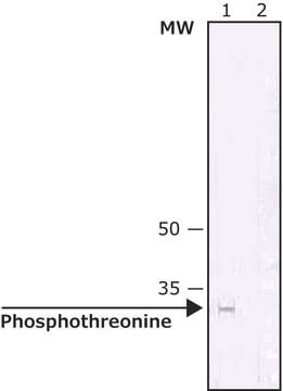
By ELISA and dot blot, the antibody reacts specifically with phosphorylated serine, both as free amino acid or conjugated to carriers such as BSA or KLH. No cross-reactivity is observed with non-phosphorylated serine, phosphothreonine, phosphotyrosine, AMP or ATP. This antibody has been used in immunoblotting for the localization of some phosphoserine-containing proteins. Certain proteins known to contain phosphorylated serine may not be recognized by this antibody due to steric hindrance of the recognition site.

Immunogen

phosphoserine conjugated to Keyhole Limpet Hemocyanin (KLH).

Application

Anti-Phosphoserine antibody may be used for indirect ELISA at a working dilution of 1:4000. For immunoblotting using rat brain cortex extracts, a working dilution of 1:500-1:1100 may be used. The antibody was used for immunoblotting to detect proteins phosphorylated at serine in stem extracts of Arabidopsis thalliana at a working dilution of 1:250.
Applications in which this antibody has been used successfully, and the associated peer-reviewed papers, are given below.
Western Blotting (1 paper)
Western blotting following immunoprecipitation (1 paper)
Monoclonal Anti-Phosphoserine antibody produced in mouse has been used in western blotting.

Biochem/physiol Actions

Phosphoserine (PS) can be used as a template to detect cancer antigen 125 (CA 125).

Disclaimer

Unless otherwise stated in our catalog or other company documentation accompanying the product(s), our products are intended for research use only and are not to be used for any other purpose, which includes but is not limited to, unauthorized commercial uses, in vitro diagnostic uses, ex vivo or in vivo therapeutic uses or any type of consumption or application to humans or animals.

Not finding the right product?  

Try our Product Selector Tool.

related product

Product No.
Description
Pricing

Storage Class

12 - Non Combustible Liquids

wgk_germany

WGK 2

flash_point_f

Not applicable

flash_point_c

Not applicable


Certificates of Analysis (COA)

Search for Certificates of Analysis (COA) by entering the products Lot/Batch Number. Lot and Batch Numbers can be found on a product’s label following the words ‘Lot’ or ‘Batch’.

Already Own This Product?

Find documentation for the products that you have recently purchased in the Document Library.

Visit the Document Library

Identification of cellulose synthase AtCesA7 (IRX3) in vivo phosphorylation sites?a potential role in regulating protein degradation
Taylor NG
Plant Molecular Biology, 64(1-2), 161-171 (2007)
Sapan J Patel et al.
Journal of cellular and molecular medicine, 21(3), 456-466 (2016-09-30)
B-cell novel protein-1 (BCNP1) or Family member of 129C (FAM129C) was identified as a B-cell-specific plasma-membrane protein. Bioinformatics analysis predicted that BCNP1 might be heavily phosphorylated. The BCNP1 protein contains a pleckstrin homology (PH) domain, two proline-rich (PR) regions and
Vinculin but not alpha-actinin is a target of PKC phosphorylation during junctional assembly induced by calcium
Perez-MM, et al.
Journal of Cell Science, 111(23), 3563-3571 (1998)
Pierrick Dudognon et al.
FEBS letters, 561(1-3), 44-50 (2004-03-12)
Endoplasmic reticulum (ER)-to-Golgi transport is blocked in mammalian cells during mitosis; however, the mechanism underlying this blockade remains unknown. Since COPII proteins are involved in this transport pathway, we investigated at the biochemical level post-translational modifications of COPII components during
Neil G Taylor
Plant molecular biology, 64(1-2), 161-171 (2007-04-12)
Cellulose is central to plant development and is synthesised at the plasma membrane by an organised protein complex that contains three different cellulose synthase proteins. The ordered assembly of these three catalytic subunits is essential for normal cellulose synthesis. The

Our team of scientists has experience in all areas of research including Life Science, Material Science, Chemical Synthesis, Chromatography, Analytical and many others.

Contact Technical Service