Skip to Content
MilliporeSigma
All Photos(2)

Key Documents

SAB4200681

Sigma-Aldrich

Anti-Green Fluorescent Protein (GFP) antibody, Mouse monoclonal

clone GFP-20, purified from hybridoma cell culture

Synonym(s):

Monoclonal Anti-Green Fluorescent Protein (GFP) antibody produced in mouse, green fluorescent protein

Sign Into View Organizational & Contract Pricing


About This Item

UNSPSC Code:
12352203
NACRES:
NA.46

biological source

mouse

Quality Level

antibody form

purified antibody

antibody product type

primary antibodies

clone

GFP-20, monoclonal

form

buffered aqueous solution

mol wt

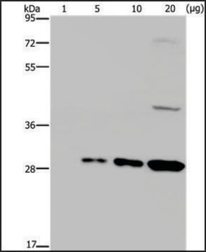
antigen 27 kDa

species reactivity

rat, human

concentration

~1 mg/mL

technique(s)

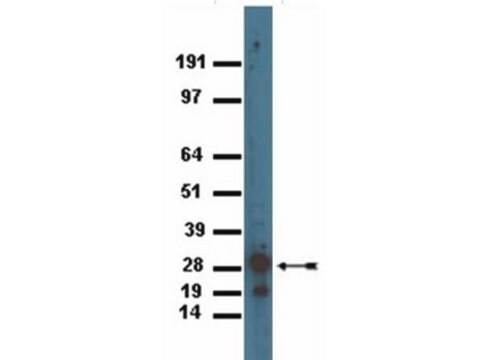
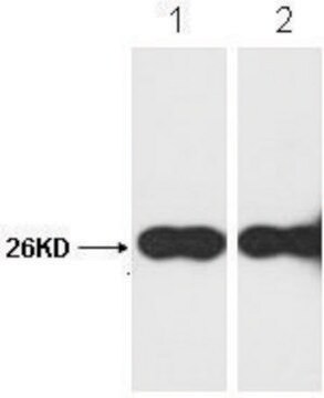
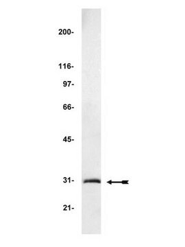
immunoblotting: 1-2 μg/mL using whole extract of human HEK-293T cells over-expressing GFP tagged fusion protein.
immunoprecipitation (IP): 5-10 μg using whole extract of human HEK-293T cells over-expressing GFP tagged fusion protein.

isotype

IgG1

shipped in

dry ice

storage temp.

−20°C

target post-translational modification

unmodified

General description

Monoclonal anti-green fluorescent protein (GFP) (mouse IgG1 isotype) is derived from the GFP-20 hybridoma produced by the fusion of mouse myeloma cells and splenocytes from mouse BALB/c mice immunized with a GFP tagged fusion protein.GFP is a 27kDa protein, derived from the bioluminescent jellyfish Aequorea victoria, in which light is produced when energy is transferred from the Ca2+-activated photoprotein aequorin to GFP.

Specificity

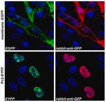
Monoclonal Anti-Green Fluorescent Protein (GFP) recognizes N-terminal and C-terminal GFP (27 kDa) tagged fusion proteins. The antibody reacts with fusion proteins expressed by prokaryotes expression vectors.

Immunogen

GFP tagged fusion protein

Application

Monoclonal Anti-Green Fluorescent Protein (GFP) antibody produced in mouse may be used in:
  • immunoblotting
  • immunoprecipitation
  • dot blot
  • enzyme-linked immunosorbent assay (ELISA)
  • scanning electron microscope (SEM)

Biochem/physiol Actions

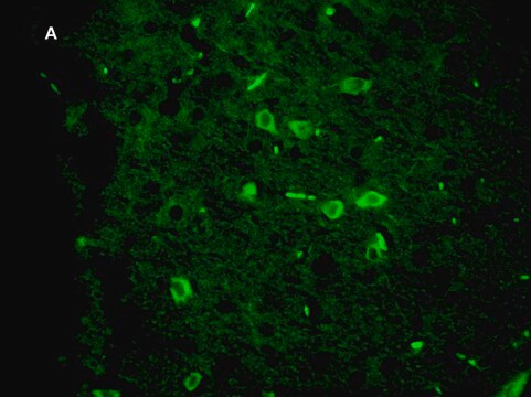
GFP (green fluorescence protein) is a reporter molecule which is used for checking gene expression and protein localization. GFP emits green light when it is excited with UV/blue light. The GFP fluorescence remains stable and can be detected non-invasively in living cells. GFP is considered as a unique tool to monitor dynamic processes in several living cells or organisms. When expressed in either eukaryotic or prokaryotic cells and illuminated by blue or UV light, GFP yields a bright green fluorescence.

Physical form

a solution in 0.01 M phosphate buffered saline pH 7.4, containing 15 mM sodium azide

Storage and Stability

For continuous use, store at 28 °C for up to one month. For extended storage, freeze in working aliquots. Repeated freezing and thawing is not recommended. If slight turbidity occurs upon prolonged storage, clarify the solution by centrifugation before use. Working dilution samples should be discarded if not used within 12 hours.

Disclaimer

Unless otherwise stated in our catalog or other company documentation accompanying the product(s), our products are intended for research use only and are not to be used for any other purpose, which includes but is not limited to, unauthorized commercial uses, in vitro diagnostic uses, ex vivo or in vivo therapeutic uses or any type of consumption or application to humans or animals.

Not finding the right product?  

Try our Product Selector Tool.

Storage Class

10 - Combustible liquids

flash_point_f

Not applicable

flash_point_c

Not applicable


Certificates of Analysis (COA)

Search for Certificates of Analysis (COA) by entering the products Lot/Batch Number. Lot and Batch Numbers can be found on a product’s label following the words ‘Lot’ or ‘Batch’.

Already Own This Product?

Find documentation for the products that you have recently purchased in the Document Library.

Visit the Document Library

Ilenia Meloni et al.
Scientific reports, 10(1), 17614-17614 (2020-10-21)
Invertebrates such as Drosophila melanogaster have proven to be a valuable model organism for studies of the nervous system. In order to control neuronal activity, optogenetics has evolved as a powerful technique enabling non-invasive stimulation using light. This requires light
Sven Dannhäuser et al.
eLife, 9 (2020-10-01)
Adhesion-type GPCRs (aGPCRs) participate in a vast range of physiological processes. Their frequent association with mechanosensitive functions suggests that processing of mechanical stimuli may be a common feature of this receptor family. Previously, we reported that the Drosophila aGPCR CIRL
The Green Fluorescent Protein
Tsien RY
Annual Review of Biochemistry, 67, 509-544 (1998)
Green Fluorescent Protein as a Reporter for Macromolecular Localization in Bacterial Cells
Margolin W
Methods, 20, 62-72 (2000)
Expression and Detection of Green Fluorescent Protein (GFP)
Kain SR and Kitts P.
Methods in Molecular Biology, 63, 305-324 (1997)

Our team of scientists has experience in all areas of research including Life Science, Material Science, Chemical Synthesis, Chromatography, Analytical and many others.

Contact Technical Service