Skip to Content
MilliporeSigma
All Photos(1)

Key Documents

SRP3324

Sigma-Aldrich

Thymosin β4 human

recombinant, expressed in E. coli, ≥95% (SDS-PAGE), ≥95% (HPLC)

Synonym(s):

Hematopoietic system regulatory peptide, Seraspenide, T-4

Sign Into View Organizational & Contract Pricing


About This Item

UNSPSC Code:
12352202
NACRES:
NA.32

biological source

human

recombinant

expressed in E. coli

assay

≥95% (HPLC)
≥95% (SDS-PAGE)

form

lyophilized

potency

0.5-10 μg/mL

mol wt

5.2 kDa

packaging

pkg of 100 μg

impurities

endotoxin, tested

UniProt accession no.

shipped in

wet ice

storage temp.

−20°C

Gene Information

human ... TMSB4X(7114)

General description

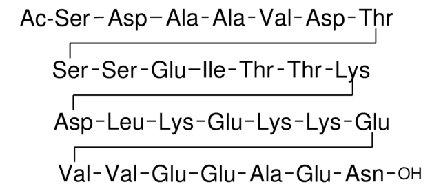
Thymosin-β4 is a small, actin-sequestering protein belonging to the thymosin-β family that is found at high concentrations within the spleen, thymus, and peritoneal macrophages, where it is most notably responsible for the organization of cytoskeletal structure. Commonly found at significant quantities within the brain, lungs, liver, kidneys, testes, and heart, Thymosin-β4 has also been shown to be synthesized by cells unrelated to the reticuloendothelial system, such as myoblasts and fibroblasts, and expressed at irregular levels by several hemopoietic cell lines, malignant lymphoid cells and myeloma cells. The gene is mapped to human chromosome Xq21-22. Recombinant Human Thymosin-β4 is a 5.2kDa glycoprotein containing 45 amino acid residues.

Biochem/physiol Actions

In mammalian tissues, thymosin-β4 protein acts as a modulator for the polymerization/depolymerization of actin through the formation of a 1:1 complex with the monomer G (globular)-actin, and inhibits actin′s polymerization to form F (filamentous) actin, which together with other proteins binds microfilaments to construct the cytoskeleton. In addition to regulating actin polymerization, research has also found thymosin-β4 to stimulate the secretion of hypothalamic luteinizing hormone-releasing hormone and luteinizing hormone, inhibit the migration of peritoneal macrophages, induce phenotypic changes in T cell lines during early host defense mechanisms, and inhibit the progression of hematopoietic pluripotent stem cells into the S-phase. It also enhances angiogenesis, endothelial cell migration and adhesion and tubule formation. Thymosin-β4 also participates in wound healing by reducing inflammation and by exhibiting anti-fibrotic effects. It also controls the migration of cancer cells.

Physical form

Lyophilized with no additives.

Reconstitution

Centrifuge the vial prior to opening. Reconstitute in water to a concentration of 0.1-1.0 mg/ml. Do not vortex. This solution can be stored at 2-8 °C for up to 1 week. For extended storage, it is recommended to further dilute in a buffer containing a carrier protein (example 0.1% BSA) and store in working aliquots at -20 °C to -80 °C.

Storage Class

11 - Combustible Solids

wgk_germany

WGK 3

flash_point_f

Not applicable

flash_point_c

Not applicable


Certificates of Analysis (COA)

Search for Certificates of Analysis (COA) by entering the products Lot/Batch Number. Lot and Batch Numbers can be found on a product’s label following the words ‘Lot’ or ‘Batch’.

Already Own This Product?

Find documentation for the products that you have recently purchased in the Document Library.

Visit the Document Library

beta-Thymosins, small acidic peptides with multiple functions.
Huff T, et al.
The International Journal of Biochemistry & Cell Biology, 33, 205-220 (2001)
Thymosin beta4: actin-sequestering protein moonlights to repair injured tissues.
Goldstein AL, et al.
Trends in Molecular Medicine, 11, 421-429 (2005)
Gender, neuroendocrine-immune interactions and neuron-glial plasticity. Role of luteinizing hormone-releasing hormone (LHRH).
Marchetti B, et al.
Annals of the New York Academy of Sciences, 917, 678-709 (2000)
The actin binding site on thymosin beta4 promotes angiogenesis.
Philp D, et al.
Faseb Journal, 17, 2103-2105 (2003)
Alexander Belyy et al.
Nature communications, 12(1), 6628-6628 (2021-11-18)
Bacterial human pathogens secrete initially inactive nucleotidyl cyclases that become potent enzymes by binding to actin inside eukaryotic host cells. The underlying molecular mechanism of this activation is, however, unclear. Here, we report structures of ExoY from Pseudomonas aeruginosa and

Our team of scientists has experience in all areas of research including Life Science, Material Science, Chemical Synthesis, Chromatography, Analytical and many others.

Contact Technical Service