MAB4197
Anti-Topoisomerase II Antibody, clone KiS1
clone KiS1, Chemicon®, from mouse
別名:
Ki-S1
ログイン組織・契約価格を表示する
すべての画像(1)
About This Item
UNSPSCコード:
12352203
eCl@ss:
32160702
NACRES:
NA.41
クローン:
KiS1, monoclonal
application:
FACS
ICC
IHC (p)
WB
ICC
IHC (p)
WB
化学種の反応性:
human
テクニック:
flow cytometry: suitable
immunocytochemistry: suitable
immunohistochemistry (formalin-fixed, paraffin-embedded sections): suitable
western blot: suitable
immunocytochemistry: suitable
immunohistochemistry (formalin-fixed, paraffin-embedded sections): suitable
western blot: suitable
citations:
21
おすすめの製品
由来生物
mouse
品質水準
抗体製品の状態
purified immunoglobulin
クローン
KiS1, monoclonal
化学種の反応性
human
メーカー/製品名
Chemicon®
テクニック
flow cytometry: suitable
immunocytochemistry: suitable
immunohistochemistry (formalin-fixed, paraffin-embedded sections): suitable
western blot: suitable
アイソタイプ
IgG2a
NCBIアクセッション番号
UniProtアクセッション番号
輸送温度
wet ice
ターゲットの翻訳後修飾
unmodified
遺伝子情報
human ... TOP2A(7153)
詳細
Topoisomerase II β (M.W 180 kDa) plays important roles in synthesis and transcription of DNA as well as chromosomal segregation during mitosis. It is present ubiquitously in normal cells and is upregulated in tumors and proliferating cells. Topoisomerase II β is also implicated in drug resistance of tumor cells.
特異性
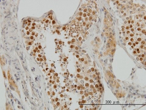
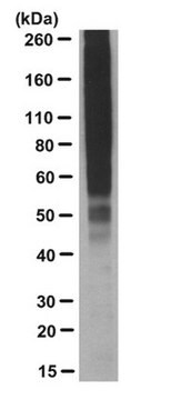
In Immunoprecipitation and Western blot experiments the Ki-S1 antibody recognizes a major protein of 170 kD which was identified as the a isoform of topoisomerase II (Boege et al., 1995; Kreipe et al., 1993). In addition, it has been shown that the Ki-S1 antibody recognizes a carboxyterminal a-isoenzyme specific epitope missing in topoisomerase IIb (Boege et al., 1995). In immunohistochemistry the Ki-S1 antibody shows strong nuclear staining only in proliferating cells. The epitope recognized by the antibody is also detectable in paraffin-embedded tissue sections (Kreipe et al., 1993). Accordingly, it has been shown that the expression of topoisomerase IIa is strongly restricted to proliferating cells (Tsutsui et al., 1993). The topoisomerase IIa antigen is preferentially expressed during G 1 , S, G 2 and M phase of the cell cycle, while resting, non-cycling cells (G 0 phase) lack topoisomerase IIa. In addition, constantly proliferating cells (e.g. cell lines) react positively to topoisomerase IIa during the entire cell-cycle. This specificity of Ki-S1 antibody for proliferating cells might make it a useful tool for determination of the proliferative fraction in solid tumors such as mammary carcinomas (Kreipe et al., 1993; Sampson et al., 1992; Camplejohn et al., 1993; Kreipe et al., 1993; Rasbridge et al., 1994) and gangliomas (Wolf et al., 1994).
アプリケーション
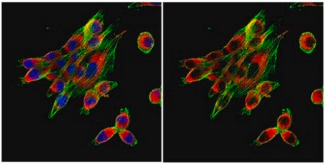
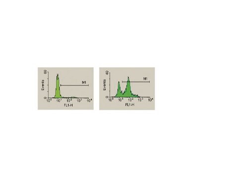
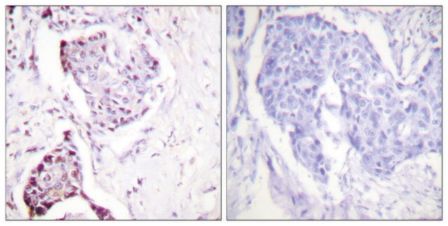
Anti-Topoisomerase II Antibody, clone KiS1 is a Mouse Monoclonal Antibody for detection of Topoisomerase II also known as Ki-S1 & has been validated in FC, WB, ICC, IHC, IHC(P).
Western blot: 1-10μg/ml Immunoprecipitaion
Immunocytochemistry: 5-10 μg/ml Immunohistochemistry: 5-10 μg/ml
Flow cytometry
Optimal working dilutions must be determined by end user.
Immunocytochemistry: 5-10 μg/ml Immunohistochemistry: 5-10 μg/ml
Flow cytometry
Optimal working dilutions must be determined by end user.
関連事項
Replaces: MABE519
物理的形状
Format: Purified
保管および安定性
Maintain at 2-8°C in undiluted aliquots for up to 6 months.Avoid repeat freeze/thaw cycles.
その他情報
Concentration: Please refer to the Certificate of Analysis for the lot-specific concentration.
法的情報
CHEMICON is a registered trademark of Merck KGaA, Darmstadt, Germany
保管分類コード
12 - Non Combustible Liquids
WGK
WGK 2
引火点(°F)
Not applicable
引火点(℃)
Not applicable
適用法令
試験研究用途を考慮した関連法令を主に挙げております。化学物質以外については、一部の情報のみ提供しています。 製品を安全かつ合法的に使用することは、使用者の義務です。最新情報により修正される場合があります。WEBの反映には時間を要することがあるため、適宜SDSをご参照ください。
Jan Code
MAB4197:
試験成績書(COA)
製品のロット番号・バッチ番号を入力して、試験成績書(COA) を検索できます。ロット番号・バッチ番号は、製品ラベルに「Lot」または「Batch」に続いて記載されています。
Ki-S1, a novel proliferative marker: flow cytometric assessment of staining in human breast carcinoma cells.
Camplejohn, R S, et al.
British Journal of Cancer, 67, 657-662 (1993)
Kendra Sweet et al.
Clinical cancer research : an official journal of the American Association for Cancer Research, 26(1), 54-60 (2019-10-23)
Induction chemotherapy results in complete remission (CR) rates of 20% to 50% among patients with poor-risk AML. Selinexor is an oral selective inhibitor of nuclear export with promising single-agent activity. By inhibiting the primary export protein, XPO1, selinexor localizes and
The effects of chemotherapy on morphology, cellular proliferation, apoptosis and oncoprotein expression in primary breast carcinoma.
Rasbridge, S A, et al.
British Journal of Cancer, 70, 335-341 (1994)
N I Valkov et al.
British journal of haematology, 108(2), 331-345 (2000-02-26)
The resistance of several leukaemic and myeloma cell lines (CCRF, L1210, HL-60, KG-1a and RPMI 8226) to VP-16 was found to increase with cell density and to be maximal (3.5- to 39-fold) in plateau phase cell cultures, as measured by
Ganglioglioma: a detailed histopathological and immunohistochemical analysis of 61 cases.
Wolf, H K, et al.
Acta neuropathologica, 88, 166-173 (1994)
ライフサイエンス、有機合成、材料科学、クロマトグラフィー、分析など、あらゆる分野の研究に経験のあるメンバーがおります。.
製品に関するお問い合わせはこちら(テクニカルサービス)
