추천 제품
생물학적 소스
mouse
Quality Level
항체 형태
purified immunoglobulin
항체 생산 유형
primary antibodies
클론
25F9, monoclonal
종 반응성
human
제조업체/상표
Chemicon®
기술
flow cytometry: suitable
immunocytochemistry: suitable
immunohistochemistry: suitable
동형
IgG1
배송 상태
wet ice
타겟 번역 후 변형
unmodified
일반 설명
ANTIGEN DISTRIBUTION:
Isolated cells: Absent on freshly isolated monocytes and other blood cells; present on 40-50% of human monocytes after 6-7 day culture, also positive on some melanoma and carcinoma cell lines. Tissue sections: Kupffer cells, histiocytes (skin), macrophages of the thymus, in the germinal centers of lymph nodes and spleen, in mamma carcinoma, melanoma, osteocarcinoma and gastric cancer; excema, sarcoidosis, BCG granuloma; synovial lining cells, tuberculoid leprosy; no expression in lepramatous leprosy.
Isolated cells: Absent on freshly isolated monocytes and other blood cells; present on 40-50% of human monocytes after 6-7 day culture, also positive on some melanoma and carcinoma cell lines. Tissue sections: Kupffer cells, histiocytes (skin), macrophages of the thymus, in the germinal centers of lymph nodes and spleen, in mamma carcinoma, melanoma, osteocarcinoma and gastric cancer; excema, sarcoidosis, BCG granuloma; synovial lining cells, tuberculoid leprosy; no expression in lepramatous leprosy.
특이성
Human late stage inflammatory macrophages. The monoclonal is suitable for use on BAL (bronchial lavage fluids) and other lavages. The antigen recognized by MAB1569 is an 86 kDa protein (unreduced conditions), probably a glycoprotein on the cell surface and within the cytoplasm of mature macrophages. It is stable to formaldehyde fixation and paraffin embedding. Enzyme digestion is recommended. The antibody has also been reported to recognize microglia stem cells (X International Congress of Neuropathology, Stockholm, le 11 September,1986.PosterA-701).
Antigen distribution: absent from freshly isolated monocytes and other blood cells; present on 40-50% of human monocytes after 6-7 days in culture, also positive on some melanoma and carcinoma lines. In tissue sections, clone identifies Kupffer cells, histiocytes (skin), macrophages of the thymus, in te germinal centers of lymph nodes and spleen, in mamma carcinoma, melanoma, osteocarcinoma and gastric cancer; excema, sarcoidosis, BCG granuloma;synovial lining cells, tuberculoid leprosy, however no expression in lepramatous leprosy.
Species reactivity is seen in human, rhesus monkey, and pig, other species not tested.
애플리케이션
Anti-Microglia Antibody, clone 25F9 detects level of Microglia & has been published & validated for use in FC, IC, IH.
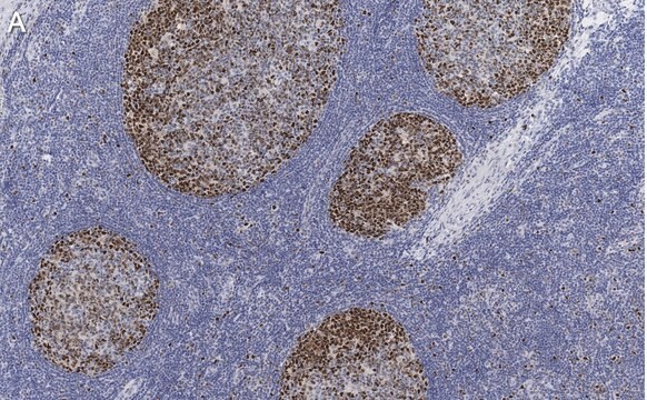
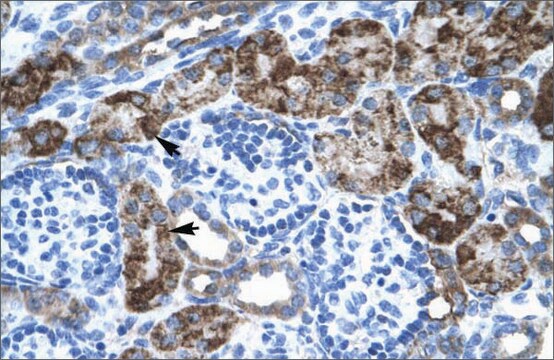
Immunohistochemistry: 1:50-1:200 using human tonsil cryosection. The antibody is reactive on formalin fixed / paraffin sections. Enzyme digestion (proteinase pretreatement) is recommended.
Immunocytochemistry
Optimal working dilutions must be determined by the end user.
Immunocytochemistry
Optimal working dilutions must be determined by the end user.
물리적 형태
Format: Purified
Purified immunoglobulin from culture supernatant. Lyophilized from PBS, pH 7.2 containing 5 mg/mL BSA and 0.09% sodium azide. Reconstitute with 500 μL of sterile distilled water.
저장 및 안정성
Maintain lyophilized material at 2-8°C for up to 6 months. After reconstitution maintain at -70°C for up to 6 months. Avoid repeated freeze/thaw cycles.
법적 정보
CHEMICON is a registered trademark of Merck KGaA, Darmstadt, Germany
적합한 제품을 찾을 수 없으신가요?
당사의 제품 선택기 도구.을(를) 시도해 보세요.
신호어
Warning
유해 및 위험 성명서
Hazard Classifications
Acute Tox. 4 Dermal - Acute Tox. 4 Inhalation - Aquatic Chronic 3
Storage Class Code
11 - Combustible Solids
WGK
WGK 3
시험 성적서(COA)
제품의 로트/배치 번호를 입력하여 시험 성적서(COA)을 검색하십시오. 로트 및 배치 번호는 제품 라벨에 있는 ‘로트’ 또는 ‘배치’라는 용어 뒤에서 찾을 수 있습니다.
Johannes Vogt et al.
EMBO molecular medicine, 8(1), 25-38 (2015-12-17)
Loss of plasticity-related gene 1 (PRG-1), which regulates synaptic phospholipid signaling, leads to hyperexcitability via increased glutamate release altering excitation/inhibition (E/I) balance in cortical networks. A recently reported SNP in prg-1 (R345T/mutPRG-1) affects ~5 million European and US citizens in a
자사의 과학자팀은 생명 과학, 재료 과학, 화학 합성, 크로마토그래피, 분석 및 기타 많은 영역을 포함한 모든 과학 분야에 경험이 있습니다..
고객지원팀으로 연락바랍니다.