추천 제품
생물학적 소스
mouse
Quality Level
100
300
항체 형태
purified immunoglobulin
항체 생산 유형
primary antibodies
클론
81 (mAB O4), monoclonal
종 반응성
rat, mouse, human, chicken
제조업체/상표
Chemicon®
기술
immunocytochemistry: suitable
immunohistochemistry: suitable
동형
IgM
적합성
not suitable for Western blot
not suitable for immunoprecipitation
배송 상태
wet ice
타겟 번역 후 변형
unmodified
특이성
Recognizes Oligodendrocyte marker O4. Also reacts with certain galactolipids in sperm (see Additional Information library for list).
애플리케이션
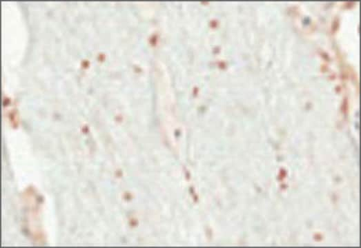
Anti-O4 Antibody, clone 81 is an antibody against O4 for use in IC, IH with more than 50 product citations.
Immunohistochemistry: 10-20 μg/mL on unfixed, shock frozen tissue.
Immunocytochemistry: 10-20 μg/mL on cells fixed with 4% paraformaldehyde.
Note: O4 is a sulfatide, which can be dissolved out of the membrane by organic solvents; acetone and methanol should not be used for fixation.
Optimal working dilutions must be determined by the end user.
Immunohistochemistry protocol
1. Prepare sections from unfixed, shock frozen tissue. The sections should be 4-5 μm thick. Place the sections on microscope slides.
2. Wash the slide three times for 5 min. each in PBS at room temperature.
3. Block the non-specific binding sites by incubating the sections in a humid chamber with 5% FCS at room temperature for 30 minutes.
4. Wash the slides as described in step 2.
5. Cover the sections with a sufficient amount of MAB345 (10-20 μg/mL in PBS) and incubate in a humid chamber at 37°C for one hour.
6. Wash the slides briefly three times with PBS. Carefully dry around the area to be stained.
7. Cover the sections with a sufficient amount of anti-mouse IgM-fluorescein* solution and incubate in a humid chamber at 37°C for one hour.
8. Wash the slides as described in step 6.
9. Cover the sections with a suitable embedding medium, cover with a cover slip, and examine by fluorescence microscopy.
*HRP or ABC can also be used.
Optimal results can be obtained by titrating the primary and secondary antibodies
Immunocytochemistry
1. Fix the preparations with 4% paraformaldehyde (in PBS) at room temperature for 10 minutes. O4 is a sulfatide which can be dissolved out of the membrane by organic solvents; acetone and methanol should not be used for fixation.
2. Wash the slide three times for 5 min. each in PBS at room temperature.
3. Block the non-specific binding sites by incubating the sections in a human chamber with 5% FCS at room temperature for 30 minutes.
4. Wash the slides as described in step 2.
5. Cover the sections with a sufficient amount of MAB345 (10-20 μg/mL in PBS) and incubate in a humid chamber at 37°C for one hour.
6. Wash the slides briefly three times with PBS. Carefully dry around the area to be stained.
7. Cover the sections with a sufficient amount of anti-mouse IgM-fluorescein solution and incubate in a humid chamber at 37°C for one hour.
8. Wash the slides as described in step 6.
9. Cover the sections with a suitable embedding medium, cover with a cover slip, and examine by fluorescence microscopy.
Note: Do not allow the preparations to dry out during staining.
물리적 형태
Format: Purified
Purified immunoglobulin in 0.05M Potassium phosphate buffer, pH 8.0 with 0.3M NaCl and 0.05% sodium azide.
분석 메모
Control
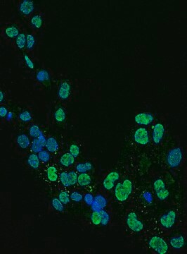
Rat cortical stem cells or day 3 cell cultures of brains from mouse embryos
Rat cortical stem cells or day 3 cell cultures of brains from mouse embryos
기타 정보
Concentration: Varies, see lot specific CoA
법적 정보
CHEMICON is a registered trademark of Merck KGaA, Darmstadt, Germany
적합한 제품을 찾을 수 없으신가요?
당사의 제품 선택기 도구.을(를) 시도해 보세요.
Storage Class Code
10 - Combustible liquids
WGK
WGK 2
시험 성적서(COA)
제품의 로트/배치 번호를 입력하여 시험 성적서(COA)을 검색하십시오. 로트 및 배치 번호는 제품 라벨에 있는 ‘로트’ 또는 ‘배치’라는 용어 뒤에서 찾을 수 있습니다.
Systemic injection of neural stem/progenitor cells in mice with chronic EAE.
Donega, M; Giusto, E; Cossetti, C; Schaeffer, J; Pluchino, S
Journal of Visualized Experiments null
Marc-André Mouthon et al.
Journal of neurochemistry, 99(3), 807-817 (2006-08-24)
Developing and adult forebrains contain neural stem cells (NSCs) but no marker is available to highly purify them. When analysed by flow cytometry, stem cells from various tissues are enriched in a 'side population' (SP) characterized by the exclusion of
Yifat Amir-Levy et al.
Multiple sclerosis international, 2014, 926134-926134 (2015-01-23)
Background. The neural stem cells (NSCs) migrate to the damaged sites in multiple sclerosis (MS) and in experimental autoimmune encephalomyelitis (EAE). However, the differentiation into neurons or oligodendrocytes is blocked. Epidermal growth factor (EGF) stimulates NSC proliferation and mobilization to
Differentiation of human breast-milk stem cells to neural stem cells and neurons.
Hosseini, SM; Talaei-Khozani, T; Sani, M; Owrangi, B
Neurology research international null
Yudong Liu et al.
Cell communication and signaling : CCS, 22(1), 155-155 (2024-03-01)
Vascular endothelial cells are pivotal in the pathophysiological progression following spinal cord injury (SCI). The UTX (Ubiquitously Transcribed Tetratripeptide Repeat on Chromosome X) serves as a significant regulator of endothelial cell phenotype. The manipulation of endogenous neural stem cells (NSCs)
자사의 과학자팀은 생명 과학, 재료 과학, 화학 합성, 크로마토그래피, 분석 및 기타 많은 영역을 포함한 모든 과학 분야에 경험이 있습니다..
고객지원팀으로 연락바랍니다.

