추천 제품
생물학적 소스
mouse
Quality Level
결합
ALEXA FLUOR™ 488
항체 형태
purified immunoglobulin
항체 생산 유형
primary antibodies
클론
A60, monoclonal
종 반응성
human, rat, mouse
제조업체/상표
Chemicon®
기술
immunohistochemistry: suitable
동형
IgG1
배송 상태
wet ice
타겟 번역 후 변형
unmodified
유전자 정보
human ... RBFOX3(146713)
mouse ... Rbfox3(52897)
rat ... Rbfox3(287847)
일반 설명
NeuN antibody (NEUronal Nuclei; clone A60) specifically recognizes the DNA-binding, neuron-specific protein NeuN, which is present in most CNS and PNS neuronal cell types of all vertebrates tested. NeuN protein distributions are apparently restricted to neuronal nuclei, perikarya and some proximal neuronal processes in both fetal and adult brain although, some neurons fail to be recognized by NeuN at all ages: INL retinal cells, Cajal-Retzius cells, Purkinje cells, inferior olivary and dentate nucleus neurons, and sympathetic ganglion cells are examples (Mullen et al., 1992; Wolf et al., 1996). Immunohistochemically detectable NeuN protein first appears at developmental timepoints that correspond with the withdrawal of the neuron from the cell cycle and/or with the initiation of terminal differentiation of the neuron (Mullen et al., 1992). Immunoreactivity appears around E9.5 in the mouse neural tube and is extensive throughout the developing nervous system by E12.5. Strong nuclear staining suggests a nuclear regulatory protein function; however, no evidence currently exists as to whether the NeuN protein antigen has a function in the distal cytoplasm or whether it is merely synthesized there before being transported back into the nucleus. No difference between protein isolated from purified nuclei and whole brain extract on immunoblots has been found (Mullen et al., 1992).
특이성
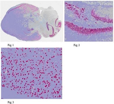
Vertebrate neuron-specific nuclear protein called NeuN (Neuronal Nuclei). MAB377X reacts with most neuronal cell types throughout the nervous system of mice including cerebellum, cerebral cortex, hippocampus, thalamus, spinal cord and neurons in the peripheral nervous system including dorsal root ganglia, sympathetic chain ganglia and enteric ganglia. The immunohistochemical staining is primarily in the nucleus of the neurons with lighter staining in the cytoplasm. The few cell types not reactive with MAB377X include Purkinje, mitral and photoreceptor cells.
Developmentally, immunoreactivity is first observed shortly after neurons have become postmitotic, no staining has been observed in proliferative zones.
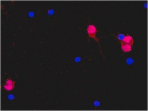
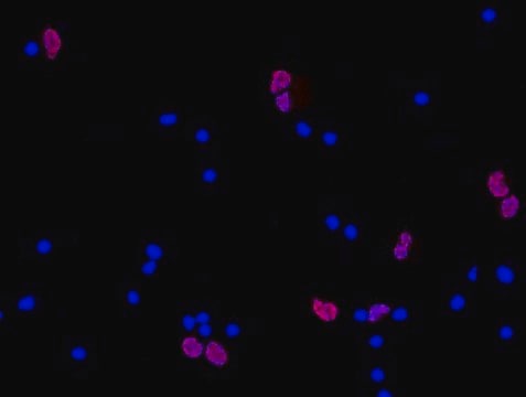
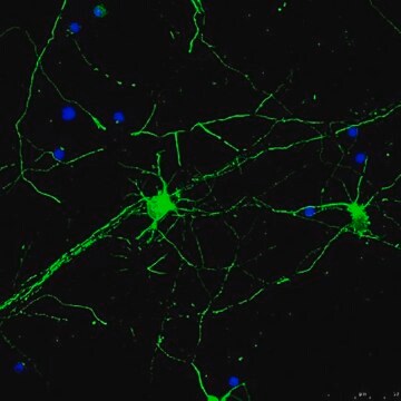
The antibody is an excellent marker for neurons in primary cultures and in retinoic acid-stimulated P19 cells. It is also useful for identifying neurons in transplants.
면역원
Purified cell nuclei from mouse brain.
애플리케이션
Anti-NeuN Antibody, clone A60, Alexa Fluor™488 conjugated is an antibody against NeuN for use in IH.
Immunohistochemistry: 1:100 on rat (paraformaldehyde fixed) and mouse (paraformaldehyde fixed, antigen retrieved) brain tissue.
Optimal working dilutions must be determined by end user.
Optimal working dilutions must be determined by end user.
Research Category
Neuroscience
Neuroscience
Research Sub Category
Neuronal & Glial Markers
Neuronal & Glial Markers
물리적 형태
Protein A purified
Purified immunoglobulin conjugated to Alexa Fluor™ 488. Liquid in Phosphate buffer with 15 mg/mL BSA as a stabilizer and 0.1% sodium azide.
저장 및 안정성
Maintain for 6 months at 2–8°C from date of shipment. Aliquot to avoid repeated freezing and thawing. For maximum recovery of product, centrifuge the original vial after thawing and prior to removing the cap.
분석 메모
Control
Brain tissue, most neuronal cell types throughout the adult nervous system
Brain tissue, most neuronal cell types throughout the adult nervous system
기타 정보
Concentration: Please refer to the Certificate of Analysis for the lot-specific concentration.
법적 정보
ALEXA FLUOR is a trademark of Life Technologies
CHEMICON is a registered trademark of Merck KGaA, Darmstadt, Germany
면책조항
Alexa Fluor™ is a registered trademark of Molecular Probes, Inc.
Unless otherwise stated in our catalog or other company documentation accompanying the product(s), our products are intended for research use only and are not to be used for any other purpose, which includes but is not limited to, unauthorized commercial uses, in vitro diagnostic uses, ex vivo or in vivo therapeutic uses or any type of consumption or application to humans or animals.
Unless otherwise stated in our catalog or other company documentation accompanying the product(s), our products are intended for research use only and are not to be used for any other purpose, which includes but is not limited to, unauthorized commercial uses, in vitro diagnostic uses, ex vivo or in vivo therapeutic uses or any type of consumption or application to humans or animals.
적합한 제품을 찾을 수 없으신가요?
당사의 제품 선택기 도구.을(를) 시도해 보세요.
Storage Class Code
12 - Non Combustible Liquids
WGK
WGK 2
Flash Point (°F)
Not applicable
Flash Point (°C)
Not applicable
시험 성적서(COA)
제품의 로트/배치 번호를 입력하여 시험 성적서(COA)을 검색하십시오. 로트 및 배치 번호는 제품 라벨에 있는 ‘로트’ 또는 ‘배치’라는 용어 뒤에서 찾을 수 있습니다.
이미 열람한 고객
The laminar development of direction selectivity in ferret visual cortex.
Clemens, JM; Ritter, NJ; Roy, A; Miller, JM; Van Hooser, SD
The Journal of Neuroscience null
Non-Gaussian diffusion imaging for enhanced contrast of brain tissue affected by ischemic stroke.
Grinberg, F; Farrher, E; Ciobanu, L; Geffroy, F; Le Bihan, D; Shah, NJ
Testing null
Gongliang Zhang et al.
Neuropharmacology, 102, 295-303 (2015-12-15)
Visceral hypersensitivity is a common characteristic in patients suffering from irritable bowel syndrome (IBS) and other disorders with visceral pain. Although the pathogenesis of visceral hypersensitivity remains speculative due to the absence of pathological changes, the long-lasting sensitization in neuronal
Jing Chen-Roetling et al.
Neuropharmacology, 56(5), 922-928 (2009-04-18)
Hemoglobin breakdown produces an iron-dependent neuronal injury after experimental CNS hemorrhage that may be attenuated by heme oxygenase (HO) inhibitors. The HO enzymes are phosphoproteins that are activated by phosphorylation in vitro. While testing the effect of kinase inhibitors in
Ali Sharma et al.
The Journal of neuroscience : the official journal of the Society for Neuroscience, 36(5), 1711-1722 (2016-02-05)
Although comprehensively described during early neuronal development, the role of DNA methylation/demethylation in neuronal lineage and subtype specification is not well understood. By studying two distinct neuronal progenitors as they differentiate to principal neurons in mouse hippocampus and striatum, we
자사의 과학자팀은 생명 과학, 재료 과학, 화학 합성, 크로마토그래피, 분석 및 기타 많은 영역을 포함한 모든 과학 분야에 경험이 있습니다..
고객지원팀으로 연락바랍니다.
