MABN153
Anti-HuC/HuD Antibody, clone 15A7.1
clone 15A7.1, from mouse
동의어(들):
ELAV-like protein 3, Hu-antigen C, HuC, Paraneoplastic cerebellar degeneration-associated antigen, Paraneoplastic limbic encephalitis antigen 21, ELAV-like protein 4, Hu-antigen D, HuD, Paraneoplastic encephalomyelitis antigen HuD
로그인조직 및 계약 가격 보기
모든 사진(2)
About This Item
UNSPSC 코드:
12352203
eCl@ss:
32160702
NACRES:
NA.41
추천 제품
생물학적 소스
mouse
Quality Level
항체 형태
purified antibody
항체 생산 유형
primary antibodies
클론
15A7.1, monoclonal
종 반응성
human, rat, mouse
기술
immunohistochemistry: suitable (paraffin)
western blot: suitable
동형
IgG2aκ
NCBI 수납 번호
배송 상태
wet ice
타겟 번역 후 변형
unmodified
유전자 정보
human ... ELAVL4(1996)
일반 설명
Hu proteins are mammalian embryonic lethal abnormal visual system (ELAV)-like neuronal RNA-binding proteins that contain three RNA recognition motifs. RNA binding protein HuC promotes neuronal development in both the central and the peripheral nervous systems. HuC antibodies are used as markers for developing neurons. HuD may play a role in neuron-specific RNA processing, and be involved in maintaining the proliferative state of cells.
면역원
KLH-conjugated linear peptide corresponding to human HuC/HuD.
애플리케이션
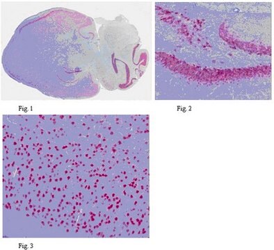
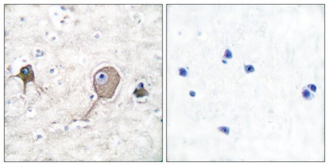
Immunohistochemistry Analysis: A 1:2,000 dilution from a representative lot detected HuC/HuD in human brain and rat hippocampus tissues.
Research Category
Neuroscience
Neuroscience
Research Sub Category
Developmental Signaling
Developmental Signaling
This HuC/HuD antibody is validated for use in WB & IHC for the detection of the HuC/HuD protein.
품질
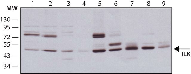
Evaluated by Western Blotting in human brain tissue lysate.
Western Blotting Analysis: 0.5 µg/mL of this antibody detected HuC/HuD in 200 µg of human brain tissue lysate.
Western Blotting Analysis: 0.5 µg/mL of this antibody detected HuC/HuD in 200 µg of human brain tissue lysate.
표적 설명
~ 40 kDa observed. An uncharacterized band may appear at ~45 kDa in some lysates.
물리적 형태
Format: Purified
Protein G Purified
Purified mouse monoclonal IgG2aκ in buffer containing 0.1 M Tris-Glycine (pH 7.4), 150 mM NaCl with 0.05% sodium azide.
저장 및 안정성
Stable for 1 year at 2-8°C from date of receipt.
분석 메모
Control
Human brain tissue lysate
Human brain tissue lysate
기타 정보
Concentration: Please refer to the Certificate of Analysis for the lot-specific concentration.
면책조항
Unless otherwise stated in our catalog or other company documentation accompanying the product(s), our products are intended for research use only and are not to be used for any other purpose, which includes but is not limited to, unauthorized commercial uses, in vitro diagnostic uses, ex vivo or in vivo therapeutic uses or any type of consumption or application to humans or animals.
적합한 제품을 찾을 수 없으신가요?
당사의 제품 선택기 도구.을(를) 시도해 보세요.
Storage Class Code
12 - Non Combustible Liquids
WGK
WGK 1
Flash Point (°F)
Not applicable
Flash Point (°C)
Not applicable
시험 성적서(COA)
제품의 로트/배치 번호를 입력하여 시험 성적서(COA)을 검색하십시오. 로트 및 배치 번호는 제품 라벨에 있는 ‘로트’ 또는 ‘배치’라는 용어 뒤에서 찾을 수 있습니다.
Karolína Liška et al.
Neuroendocrinology, 112(4), 384-398 (2021-06-11)
Circadian clocks in the hippocampus (HPC) align memory processing with appropriate time of day. Our study was aimed at ascertaining the specificity of glycogen synthase kinase 3-beta (GSK3β)- and glucocorticoid (GC)-dependent pathways in the entrainment of clocks in individual HPC
Yaxing Gui et al.
Brain pathology (Zurich, Switzerland), 30(1), 151-164 (2019-07-06)
The 18 kDa translocator protein (TSPO) is a widely used target for microglial PET imaging radioligands, but its expression in post-mortem normal and diseased human brain is not well described. We aimed at characterizing the TSPO expression in human control
Clara Muñoz-Castro et al.
Methods in molecular biology (Clifton, N.J.), 2593, 283-305 (2022-12-14)
There is a growing interest in expanding the multiplexing capability of immunohistochemistry to achieve a deeper phenotyping of various cell types in health and disease. Here, we describe a protocol of cyclic multiplex fluorescent immunohistochemistry that enables the labeling of
Joslyn Lay et al.
American journal of physiology. Gastrointestinal and liver physiology, 311(2), G252-G266 (2016-07-02)
The μ-opioid receptor (MOR) is a major regulator of gastrointestinal motility and secretion and mediates opiate-induced bowel dysfunction. Although MOR is of physiological and therapeutic importance to gut function, the cellular and subcellular distribution and regulation of MOR within the
Christine Marques et al.
Acta neuropathologica, 147(1), 56-56 (2024-03-13)
The stimulator of interferon genes (STING) pathway has been implicated in neurodegenerative diseases, including Parkinson's disease and amyotrophic lateral sclerosis (ALS). While prior studies have focused on STING within immune cells, little is known about STING within neurons. Here, we
자사의 과학자팀은 생명 과학, 재료 과학, 화학 합성, 크로마토그래피, 분석 및 기타 많은 영역을 포함한 모든 과학 분야에 경험이 있습니다..
고객지원팀으로 연락바랍니다.