
MABN24
Anti-pan-Shank Antibody, clone N23B/49
clone N23B/49, from mouse
동의어(들):
SH3 and multiple ankyrin repeat domains 2, SH3 and multiple ankyrin repeat domains protein 2, proline-rich synapse associated protein 1, cortactin SH3 domain-binding protein, Cortactin-binding protein 1, Proline-rich synapse-associated protein 1, cortact
로그인조직 및 계약 가격 보기
모든 사진(3)
About This Item
UNSPSC 코드:
12352203
eCl@ss:
32160702
NACRES:
NA.41
추천 제품


생물학적 소스
mouse
Quality Level
항체 형태
purified antibody
항체 생산 유형
primary antibodies
클론
N23B/49, monoclonal
종 반응성
mouse, human, rat
기술
immunohistochemistry: suitable (paraffin)
western blot: suitable
동형
IgG1κ
NCBI 수납 번호
UniProt 수납 번호
배송 상태
wet ice
타겟 번역 후 변형
unmodified
유전자 정보
human ... SHANK2(22941)
일반 설명
Shank1, Shank2, and Shank3 make up a family of proteins that may function as molecular scaffolds in the postsynaptic density (PSD). Shank contains multiple domains for protein-protein interaction including ankyrin repeats, an SH3 domain, a PSD-95/Dlg/ZO-1 domain, a sterile α motif domain, and a proline-rich region. Alternative splicing in the Shank family may be a mechanism that regulates the molecular structure of Shank and the spectrum of Shank-interacting proteins in the PSDs of adult and developing brain.
면역원
Recombinant protein consisting of SH3/PDZ domain of rat Shank2.
애플리케이션
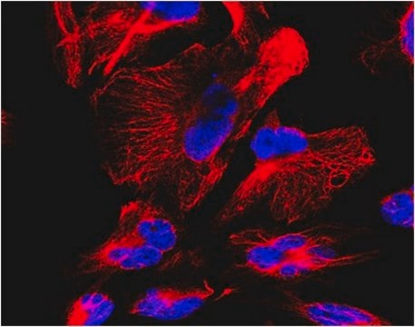
Detect pan-Shank using this Anti-pan-Shank Antibody, clone N23B/49 validated for use in WB, IH(P).
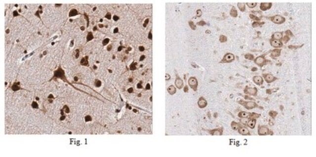
Immunohistochemistry Analysis: 1:500 dilution from a previous lot detected Shank in rat cerebellum tissue.
Western Blot Analysis: A previous lot of this antibody detected Shank in extracts of COS-1 cells transiently transfected with Shank1, Shank2 or Shank3 plasmids. Courtesy of James Trimmer, UC Davis/NIH NeuroMab Facility.
Western Blot Analysis: A previous lot of this antibody detected Shank in extracts of COS-1 cells transiently transfected with Shank1, Shank2 or Shank3 plasmids. Courtesy of James Trimmer, UC Davis/NIH NeuroMab Facility.
Research Category
Neuroscience
Neuroscience
Research Sub Category
Synapse & Synaptic Biology
Synapse & Synaptic Biology
품질
Evaluated by Western Blot in rat brain membrane tissue lysate.
Western Blot Analysis: 2 µg/mL of this antibody detected Shank on 10 µg of rat brain membrane tissue lysate.
Western Blot Analysis: 2 µg/mL of this antibody detected Shank on 10 µg of rat brain membrane tissue lysate.
표적 설명
~160 kDa observed. Other isoforms may be observed in some lysates.
물리적 형태
Format: Purified
Protein G
Purified mouse monoclonal IgG1κ in buffer containing 0.1 M Tris-Glycine (pH 7.4), 150 mM NaCl with 0.05% sodium azide.
저장 및 안정성
Stable for 1 year at 2-8°C from date of receipt.
분석 메모
Control
Rat brain membrane tissue lysate
Rat brain membrane tissue lysate
기타 정보
Concentration: Please refer to the Certificate of Analysis for the lot-specific concentration.
면책조항
Unless otherwise stated in our catalog or other company documentation accompanying the product(s), our products are intended for research use only and are not to be used for any other purpose, which includes but is not limited to, unauthorized commercial uses, in vitro diagnostic uses, ex vivo or in vivo therapeutic uses or any type of consumption or application to humans or animals.
적합한 제품을 찾을 수 없으신가요?
당사의 제품 선택기 도구.을(를) 시도해 보세요.
Storage Class Code
12 - Non Combustible Liquids
WGK
WGK 1
Flash Point (°F)
Not applicable
Flash Point (°C)
Not applicable
시험 성적서(COA)
제품의 로트/배치 번호를 입력하여 시험 성적서(COA)을 검색하십시오. 로트 및 배치 번호는 제품 라벨에 있는 ‘로트’ 또는 ‘배치’라는 용어 뒤에서 찾을 수 있습니다.
Marián Haburčák et al.
Frontiers in synaptic neuroscience, 14, 995474-995474 (2022-10-18)
The Spontaneously Hypertensive Rat (SHR) has increased sympathetic drive to the periphery that precedes and contributes to the development of high blood pressure, making it a useful model for the study of neurogenic hypertension. Comparisons to the normotensive Wistar Kyoto
Kenneth R Myers et al.
Frontiers in molecular neuroscience, 15, 1020949-1020949 (2022-10-18)
Dendritic spines are small actin-rich protrusions essential for the formation of functional circuits in the mammalian brain. During development, spines begin as dynamic filopodia-like protrusions that are then replaced by relatively stable spines containing an expanded head. Remodeling of the
Yusuke Hatanaka et al.
Scientific reports, 5, 16102-16102 (2015-11-05)
Late-onset neurodegenerative diseases are characterized by neurological symptoms and progressive neuronal death. Accumulating evidence suggests that neuronal dysfunction, rather than neuronal death, causes the symptoms of neurodegenerative diseases. However, the mechanisms underlying the dysfunction that occurs prior to cell death
자사의 과학자팀은 생명 과학, 재료 과학, 화학 합성, 크로마토그래피, 분석 및 기타 많은 영역을 포함한 모든 과학 분야에 경험이 있습니다..
고객지원팀으로 연락바랍니다.