
C2562
Monoclonal Anti-Cytokeratin, pan (Mixture) antibody produced in mouse
clone C-11+PCK-26+CY-90+KS-1A3+M20+A53-B/A2, ascites fluid
동의어(들):
Monoclonal Anti-Cytokeratin, pan (mixture), Panck Antibody, Panck Antibody - Monoclonal Anti-Cytokeratin, pan (Mixture) antibody produced in mouse
About This Item
추천 제품


생물학적 소스
mouse
Quality Level
결합
unconjugated
항체 형태
ascites fluid
항체 생산 유형
primary antibodies
클론
C-11+PCK-26+CY-90+KS-1A3+M20+A53-B/A2, monoclonal
포함
7% horse serum and 15 mM sodium azide as preservative
종 반응성
wide range
기술
immunohistochemistry (formalin-fixed, paraffin-embedded sections): suitable using protease-digested sections of human or animal tissues
immunohistochemistry (frozen sections): suitable
indirect immunofluorescence: 1:100 using protease-digested, formalin-fixed, paraffin-embedded sections of human or animal tissues
western blot: suitable
동형
IgG1/IgG2a
배송 상태
dry ice
저장 온도
−20°C
타겟 번역 후 변형
unmodified
유사한 제품을 찾으십니까? 방문 제품 비교 안내
일반 설명
특이성
면역원
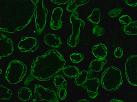
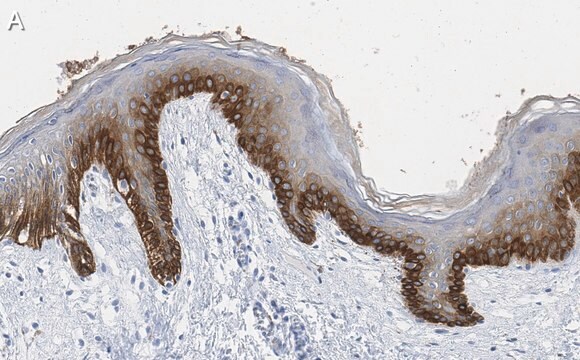
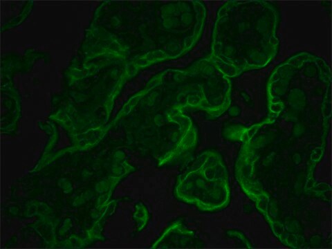
애플리케이션
- Immunohistochemistry (formalin-fixed, paraffin-embedded sections) using protease-digested sections of human or animal tissues.
- Immunohistochemistry (frozen sections).
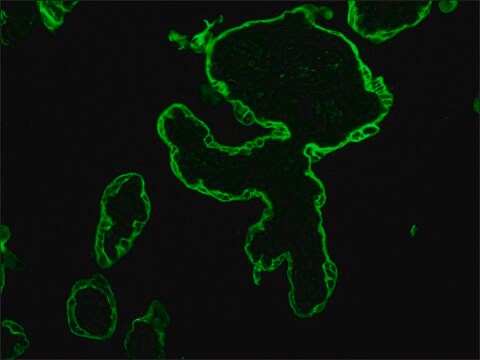
- Indirect immunofluorescence (at a working dilution of 1:100 using protease-digested, formalin-fixed, paraffin-embedded sections of human or animal tissues).
- Immunocytochemical labeling (immunofluorescence ) of cells.
- Western blotting.
생화학적/생리학적 작용
면책조항
적합한 제품을 찾을 수 없으신가요?
당사의 제품 선택기 도구.을(를) 시도해 보세요.
Storage Class Code
10 - Combustible liquids
WGK
WGK 3
가장 최신 버전 중 하나를 선택하세요:
시험 성적서(COA)
이미 열람한 고객

자사의 과학자팀은 생명 과학, 재료 과학, 화학 합성, 크로마토그래피, 분석 및 기타 많은 영역을 포함한 모든 과학 분야에 경험이 있습니다..
고객지원팀으로 연락바랍니다.