
추천 제품


생물학적 소스
mouse
Quality Level
결합
unconjugated
항체 형태
ascites fluid
항체 생산 유형
primary antibodies
클론
NOS-B1, monoclonal
분자량
antigen 150-160 kDa
포함
15 mM sodium azide
종 반응성
rat, goat, pig, human
기술
microarray: suitable
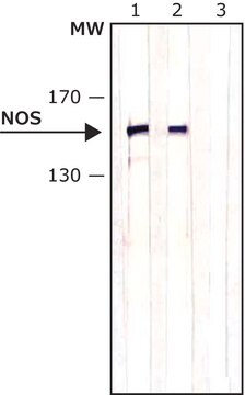
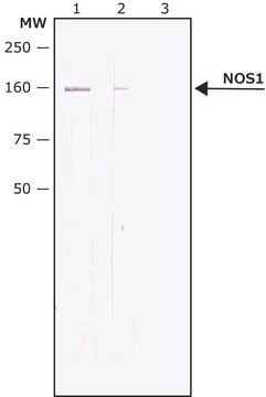
western blot: 1:3,000 using fresh rat cerebellum extract
동형
IgG1
UniProt 수납 번호
배송 상태
dry ice
저장 온도
−20°C
타겟 번역 후 변형
unmodified
유전자 정보
human ... NOS1(4842)
rat ... Nos1(24598)
일반 설명
Monoclonal Anti-Nitric Oxide Synthase-Brain (bNOS) (mouse IgG1 isotype) is derived from the NOS-B1 hybridoma produced by the fusion of mouse myeloma cells and splenocytes from immunized BALB/c mice. Nitric oxide synthase (NOS) has been localized in many different cell types. Type I NOS is found in neurons. It is a 150-160 kDa protein, also called NOS-1, neuronal NOS (nNOS), brain NOS (bNOS), cerebral NOS, constitutive NOS or Ca2+- regulated NOS (cNOS). bNOS is present also in skeletal muscle, where it is complexed with dystrophin, and is absent in Duchenne′s muscular dystrophy, which perhaps accounts for symptoms of the disease.
특이성
Monoclonal Anti-Nitric Oxide Synthase, Brain (1-181) antibody is specific for nitric oxide synthase (NOS) derived from brain (bNOS, 150-160 kDa and several breakdown products of lower M.W.). The product does not bind to NOS derived from macrophages (mNOS) and endothelial cells (eNOS). This antibody is specific for bNOS in humans, goats, pigs and rats.
면역원
recombinant neuronal NOS fragment (amino acids 1-181) from rat brain.
애플리케이션
Monoclonal Anti-Nitric Oxide Synthase, Brain (1-181) antibody produced in mouse has been used in:
- western blot analysis
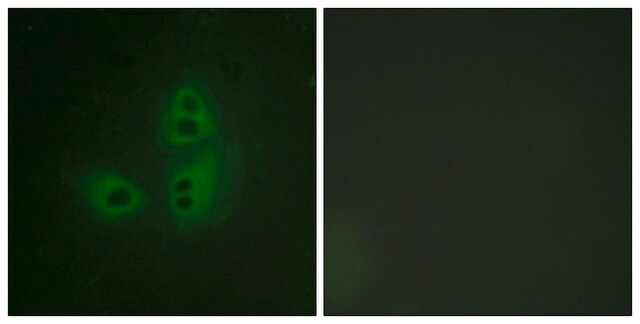
- immunocytochemistry
- double immunofluorescence
- immunofluorescence staining
- enzyme-linked immunosorbent assay (ELISA)
- dot blot immunoassay
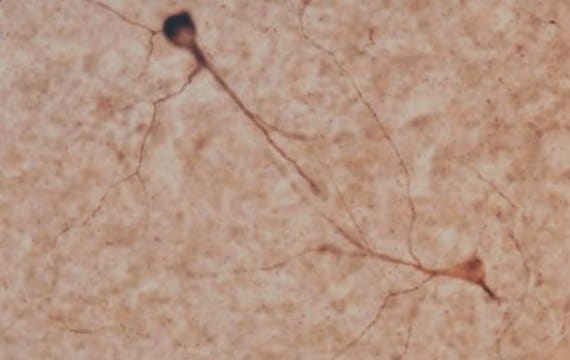
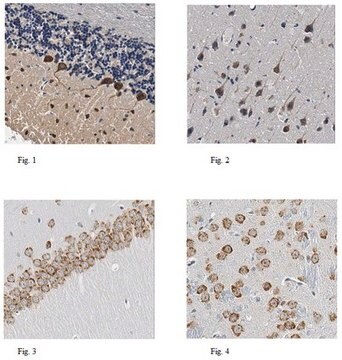
- immunohistochemical staining
생화학적/생리학적 작용
Nitric oxide synthase is an enzyme that catalyzes the formation of nitric oxide. Nitric oxide is responsible for regulating several biochemical functions such as hemostasis, neurotransmission and vascular injury response . Mutations in nitric oxide synthase gene have been associated with diabetic nephropathy.
면책조항
Unless otherwise stated in our catalog or other company documentation accompanying the product(s), our products are intended for research use only and are not to be used for any other purpose, which includes but is not limited to, unauthorized commercial uses, in vitro diagnostic uses, ex vivo or in vivo therapeutic uses or any type of consumption or application to humans or animals.
적합한 제품을 찾을 수 없으신가요?
당사의 제품 선택기 도구.을(를) 시도해 보세요.
Storage Class Code
10 - Combustible liquids
WGK
WGK 3
Flash Point (°F)
Not applicable
Flash Point (°C)
Not applicable
가장 최신 버전 중 하나를 선택하세요:
시험 성적서(COA)
Lot/Batch Number
The distribution of nitric oxide synthase in the inferior colliculus of guinea pig
Coote EJ, et al.
Neuroscience, 154(1), 218-225 (2008)
Elevated expression of CAPON and neuronal nitric oxide synthase in the sciatic nerve of rats following constriction injury
Cui Z, et al.
The Veterinary Journal, 187(3), 374-380 (2011)
The complex contribution of NOS interneurons in the physiology of cerebrovascular regulation
Duchemin S, et al.
Frontiers in Neural Circuits, 6 (2012)
Ben Coomber et al.
Frontiers in neurology, 6, 53-53 (2015-03-26)
A significant challenge in tinnitus research lies in explaining how acoustic insult leads to tinnitus in some individuals, but not others. One possibility is genetic variability in the expression and function of neuromodulators - components of neural signaling that alter
Chadd M Funk et al.
The Journal of neuroscience : the official journal of the Society for Neuroscience, 37(38), 9132-9148 (2017-08-20)
During non-rapid eye-movement (NREM) sleep, cortical and thalamic neurons oscillate every second or so between ON periods, characterized by membrane depolarization and wake-like tonic firing, and OFF periods, characterized by membrane hyperpolarization and neuronal silence. Cortical slow waves, the hallmark
자사의 과학자팀은 생명 과학, 재료 과학, 화학 합성, 크로마토그래피, 분석 및 기타 많은 영역을 포함한 모든 과학 분야에 경험이 있습니다..
고객지원팀으로 연락바랍니다.