콘텐츠로 건너뛰기
Merck
모든 사진(1)

주요 문서

O4886

Sigma-Aldrich

Monoclonal Anti-Opsin antibody produced in mouse

clone RET-P1, ascites fluid

동의어(들):

Anti-Opsin Antibody, Mouse Anti-Opsin, Opsin Detection Antibody

로그인조직 및 계약 가격 보기


About This Item

MDL number:
UNSPSC 코드:
12352203
NACRES:
NA.41
결합:
unconjugated
application:
ELISA (i)
EM
ICC
IHC (f)
RIA
WB
클론:
RET-P1, monoclonal
종 반응성:
goldfish, mouse, tiger salamander, rat, amphibian, quail, dove, duck, rabbit, bovine, human, turtle
citations:
54
기술:
electron microscopy: suitable
immunocytochemistry: suitable using cultured cells
immunohistochemistry (frozen sections): 1:10,000 using frozen sections of rat eye
indirect ELISA: suitable
radioimmunoassay: suitable
western blot: suitable

생물학적 소스

mouse

Quality Level

결합

unconjugated

항체 형태

ascites fluid

항체 생산 유형

primary antibodies

클론

RET-P1, monoclonal

분자량

antigen 39 kDa by immunoblotting (IB of rat retina produces closely spaced doublet)

포함

15 mM sodium azide

종 반응성

goldfish, mouse, tiger salamander, rat, amphibian, quail, dove, duck, rabbit, bovine, human, turtle

기술

electron microscopy: suitable
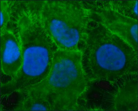
immunocytochemistry: suitable using cultured cells
immunohistochemistry (frozen sections): 1:10,000 using frozen sections of rat eye
indirect ELISA: suitable
radioimmunoassay: suitable
western blot: suitable

동형

IgG1

UniProt 수납 번호

배송 상태

dry ice

저장 온도

−20°C

타겟 번역 후 변형

unmodified

유전자 정보

human ... RHO(6010)
mouse ... Rho(212541)
rat ... Rho(24717)

일반 설명

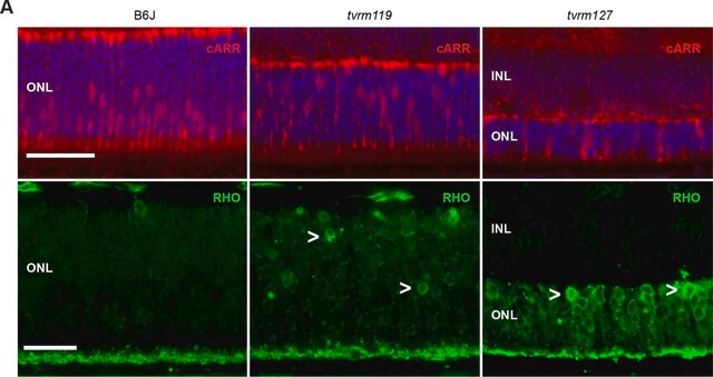
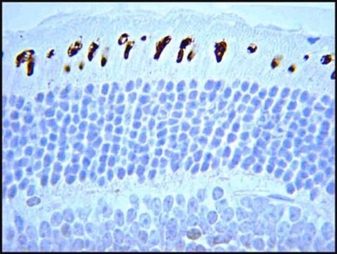
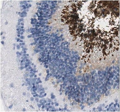
Mouse monoclonal clone RET-P1 anti-Opsin antibody recognizes an epitope located in amino acid residues 4-10 at the N-terminus of the rhodopsin molecule. In immunoblotting of rat retina, the antibody labels a closely spaced doublet of 39 kDa and less intense bands of 78 and 115 kDa, representing rhodopsin monomer and aggregates. Higher M.W. bands are stained in pituitary (251 and 288 kDa) and in hypothalamus (269 and 331 kDa) in addition to several lower M.W. bands. The product specifically labels the cell bodies, outer and inner segments (rods but not cones) of rat photoreceptor surface. It reacts with bovine, duck, amphibian, goldfish, rabbit, rat, turtle, tiger salamander, mouse, dove, quail, and human rhodopsin/opsin.
Opsin belongs to rhodopsin receptor. The gene is located on human chromosome 3q22.1. Opsin gene is mainly expressed in rod cells.
Photoreceptors are responsible for the initial step in visual processing, converting the signal of photon absorption into synaptic transmission. The visual pigment in vertebrate rods responsible for the absorption of light quanta is rhodopsin (also called opsin). Rhodopsin comprises >95% of the rod outer segments (ROS) intrinsic membrane protein and is a glycoprotein possessing two asparagine-linked oligosaccharide groups at amino acid residues 2 and 15 of the N-terminus. Light is absorbed by rhodopsin and the subsequent conformational change leads to the activation of a cyclic GMP phosphodiesterase through a specific G-protein intermediate, transducin.

면역원

rat retinal membranes.

애플리케이션

Monoclonal Anti-Opsin antibody produced in mouse has been used in immunofluorescence and histology.
Mouse monoclonal clone RET-P1 anti-Opsin antibody is used to tag opsin/rhodopsin for detection and quantitation by immunocytochemical and immunohistochemical (IHC) techniques such as immunoblotting, immunocytochemistry of cultured cells, immunohistochemistry (paraformaldehyde/glutaraldehyde-fixed, paraformaldehyde perfusion-fixed, frozen sections), immunoelectron microscopy, ELISA, competitive ELISA, and solid phase RIA. It is used as a probe to determine the presence and roles of opsin/rhodopsin in studies of rhodopsin location, functional properties and molecular mechanisms governing rod photoreceptor differentiation.

생화학적/생리학적 작용

Mutations in rhodopsin gene are linked to retinitis pigmentosa (RP) and congenital night blindness.

면책조항

Unless otherwise stated in our catalog or other company documentation accompanying the product(s), our products are intended for research use only and are not to be used for any other purpose, which includes but is not limited to, unauthorized commercial uses, in vitro diagnostic uses, ex vivo or in vivo therapeutic uses or any type of consumption or application to humans or animals.

적합한 제품을 찾을 수 없으신가요?  

당사의 제품 선택기 도구.을(를) 시도해 보세요.

Storage Class Code

10 - Combustible liquids

WGK

WGK 1

Flash Point (°F)

Not applicable

Flash Point (°C)

Not applicable

개인 보호 장비

Eyeshields, Gloves, multi-purpose combination respirator cartridge (US)


가장 최신 버전 중 하나를 선택하세요:

시험 성적서(COA)

Lot/Batch Number

적합한 버전을 찾을 수 없으신가요?

특정 버전이 필요한 경우 로트 번호나 배치 번호로 특정 인증서를 찾을 수 있습니다.

이 제품을 이미 가지고 계십니까?

문서 라이브러리에서 최근에 구매한 제품에 대한 문서를 찾아보세요.

문서 라이브러리 방문

이미 열람한 고객

Slide 1 of 1

1 of 1

Nuclear architecture of rod photoreceptor cells adapts to vision in mammalian evolution
Solovei I, et al.
Cell, 137(2), 356-368 (2009)
Sutisak Kitareewan et al.
International journal of oncology, 33(2), 397-404 (2008-07-19)
All-trans-retinoic acid (RA) treatment of acute promyelocytic leukemia (APL) cases expressing the t(15;17) product, PML/RARalpha, is a successful example of differentiation therapy. Uncovering RA target genes is of considerable interest in APL. This study comprehensively examines in APL cells transcriptional
Rhodopsin mutation G90D and a molecular mechanism for congenital night blindness
Rao VR, et al.
Nature, 367(6464), 639-639 (1994)
Autologous transplantation of RPE with partial-thickness choroid after mechanical debridement of Bruch membrane in the rabbit
Hu Y, et al.
Investigative Ophthalmology & Visual Science, 49(7), 3185-3192 (2008)
Monoclonal antibodies which recognize different cell types in the rat retina
Barnstable, C.J.
Nature, 286, 231-231 (1980)

자사의 과학자팀은 생명 과학, 재료 과학, 화학 합성, 크로마토그래피, 분석 및 기타 많은 영역을 포함한 모든 과학 분야에 경험이 있습니다..

고객지원팀으로 연락바랍니다.