크기 선택
모든 사진(2)
크기 선택
보기 변경
About This Item
실험식(Hill 표기법):
C60H70N12O13S2
Molecular Weight:
1231.40
MDL number:
UNSPSC 코드:
12352116
NACRES:
NA.32
추천 제품
제품명
Phalloidin–Tetramethylrhodamine B isothiocyanate, sequence from Amanita phalloides(synthetic: peptide sequence)
생물학적 소스
sequence from Amanita phalloides (synthetic: peptide sequence)
Quality Level
양식
solid
형광
λex 540-545 nm; λem 570-573 nm
저장 온도
−20°C
일반 설명
Phalloidin is a phallotoxin produced by death cap mushroom Amanita phalloides. It is a cyclic peptide, which interacts with actin, and this was first identified in phalloidin-poisoned rats. It is a heptapeptide, cyclic in nature, with a crosslink between tryptophan at position 6 and cysteine at position 3. The side chain of amino acid 7 (γ-δ-dihydroxyleucine) in phalloidin, is accessible to modifications, through which florescent labelled phalloidin compounds can be produced.
애플리케이션
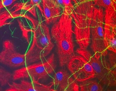
Fluorescent phallotoxin which may be used to identify filamentous actin.
Phalloidin-Tetramethylrhodamine B isothiocyanate has been used:-
Phalloidin-Tetramethylrhodamine B isothiocyanate has been used:-
- In Immunofluorescence for staining Filamentous actin (F-actin)
- To stain cells during immunocytochemical and cytochemical analysis
- To label actin microfilaments for fluorescence microscopy
생화학적/생리학적 작용
Phalloidin interacts with polymeric actin, and not oligomeric or monomeric forms. This interaction leads to highly stabilized actin filaments, which resist depolymerization and disassembly. In rats, this toxin causes death due to liver hemorrhage, and cells show abnormal actin clustering. The affinity of phalloidin to actin is not significantly altered after derivatizing florescent labelled phalloidin compounds. These compounds can be used to study actin structure and organization within eukaryotic cells.
Toxin that binds polymeric F actin, stabilizing it and interfering with the function of actin-rich structures.
기타 정보
May contain mixed isomers
관련 제품
신호어
Danger
유해 및 위험 성명서
Hazard Classifications
Acute Tox. 2 Dermal - Acute Tox. 2 Inhalation - Acute Tox. 2 Oral
Storage Class Code
6.1A - Combustible acute toxic Cat. 1 and 2 / very toxic hazardous materials
WGK
WGK 3
Flash Point (°F)
Not applicable
Flash Point (°C)
Not applicable
개인 보호 장비
Eyeshields, Faceshields, Gloves, type ABEK (EN14387) respirator filter
이미 열람한 고객
Nadav Sorek et al.
Plant physiology, 155(2), 706-720 (2010-12-09)
Prenylation primarily by geranylgeranylation is required for membrane attachment and function of type I Rho of Plants (ROPs) and Gγ proteins, while type II ROPs are attached to the plasma membrane by S-acylation. Yet, it is not known how prenylation
Emiko Hiraoka et al.
Breast cancer (Tokyo, Japan), 26(5), 581-593 (2019-03-05)
Pseudopodia are actin-rich ventral protrusions associated with cell motility and cancer cell invasion. We previously applied our established method of using excimer laser cell etching to isolate pseudopodial proteins from MDA-MB-231 breast cancer cells. We later identified 14-3-3γ as an
E Wulf et al.
Proceedings of the National Academy of Sciences of the United States of America, 76(9), 4498-4502 (1979-09-01)
A fluorescent derivative of phalloidin has been synthesized possessing high affinity to filamentous actin. This compound was used for visualization of actin-containing structures in eukaryotic nonmuscle cells. Due to its low molecular weight (1250), fixation for formaldehyde was sufficient to
Cristina Belgiovine et al.
PloS one, 5(11), e14154-e14154 (2011-01-07)
Mesenchymal and amoeboid movements are two important mechanisms adopted by cancer cells to invade the surrounding environment. Mesenchymal movement depends on extracellular matrix protease activity, amoeboid movement on the RhoA-dependent kinase ROCK. Cancer cells can switch from one mechanism to
E Langelier et al.
The journal of histochemistry and cytochemistry : official journal of the Histochemistry Society, 48(10), 1307-1320 (2000-09-16)
We investigated the structure of the chondrocyte cytoskeleton in intact tissue sections of mature bovine articular cartilage using confocal fluorescence microscopy complemented by protein extraction and immunoblotting analysis. Actin microfilaments were present inside the cell membrane as a predominantly cortical
관련 콘텐츠
Three-dimensional (3D) printing of biological tissue is rapidly becoming an integral part of tissue engineering.
자사의 과학자팀은 생명 과학, 재료 과학, 화학 합성, 크로마토그래피, 분석 및 기타 많은 영역을 포함한 모든 과학 분야에 경험이 있습니다..
고객지원팀으로 연락바랍니다.


