Skip to Content
MilliporeSigma
All Photos(3)

Documents

05-1424

Sigma-Aldrich

Anti-Endoglin Antibody, clone P3D1

clone P3D1, from mouse

Synonym(s):

CD105, Ancillary TGF-beta Receptor

Sign Into View Organizational & Contract Pricing


About This Item

UNSPSC Code:
12352203
eCl@ss:
32160702
NACRES:
NA.41

biological source

mouse

Quality Level

antibody form

purified immunoglobulin

antibody product type

primary antibodies

clone

P3D1, monoclonal

species reactivity

mouse, human

species reactivity (predicted by homology)

rat

technique(s)

ELISA: suitable
flow cytometry: suitable
immunocytochemistry: suitable
immunohistochemistry: suitable
immunoprecipitation (IP): suitable
western blot: suitable

isotype

IgG2a

NCBI accession no.

UniProt accession no.

shipped in

wet ice

target post-translational modification

unmodified

Gene Information

human ... ENG(2022)
mouse ... Eng(13805)
rat ... Eng(497010)

Related Categories

General description

Endoglin is a homodimeric integral membrane glycoprotein comprised of 95 kDa subunit. Endoglin is a component of the TGF-B receptor system that functions as an ancillary receptor influencing binding of the Transforming Growth Factor beta (TGF-β) family ligands to signaling receptors. Endoglin binds TGF-B1 and TGF-B3 with high affinity but does not bind TGF-B2. Endoglin is expressed by vascular endothelial cells and syncytiotrophoblasts of placenta and is weakly expressed by stromalfibroblasts. It is also expressed by U937 cells and activated macrophages. Endoglin expression is up regulated on activated endothelium in tissues undergoing angiogenesis, such as in tumors, or in cases of wound healing or dermal inflammation.

Specificity

Reacts with human endoglin.

Immunogen

Human resting umbilical vein endothelial cells.

Application

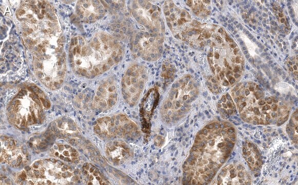
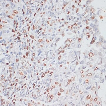
Anti-Endoglin Antibody, clone P3D1 is an antibody against Endoglin for use in WB, IH, IC, IP, FC, ELISA.
Immunohistochemistry: For use on acetone or paraformaldehyde fixed tissue.

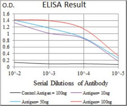
ELISA: A previous lot of this antibody was used in ELISA.

Immunoprecipitation: A previous lot of this antibody was used in IP.

Immunocytochemistry: A previous lot of this antibody was used in IC.

Flow Cytometry: A previous lot of this antibody was used in FC.

Optimal working dilutions must be determined by the end user.

Research Category
Signaling
Research Sub Category
Growth Factors & Receptors

Quality

Routinely evaluated by Western Blot on Jurkat lysates.

Western Blot Analysis: 1:500 dilution of this lot detected ENDOGLIN on 10 μg of Jurkat lysates.

Target description

~71 kDa

Linkage

Replaces: MAB2152

Physical form

Format: Purified
Purified
Purified mouse monoclonal IgG2a in 0.02M phosphate buffer, 0.25M sodium chloride, 0.1% sodium azide, pH 7.6

Storage and Stability

Stable at 2-8°C for up to 1 year from date of receipt.

Analysis Note

Control
Jurkat cell lysate

Other Notes

Concentration: Please refer to the Certificate of Analysis for the lot-specific concentration.

Disclaimer

Unless otherwise stated in our catalog or other company documentation accompanying the product(s), our products are intended for research use only and are not to be used for any other purpose, which includes but is not limited to, unauthorized commercial uses, in vitro diagnostic uses, ex vivo or in vivo therapeutic uses or any type of consumption or application to humans or animals.

wgk_germany

WGK 2

flash_point_f

Not applicable

flash_point_c

Not applicable


Certificates of Analysis (COA)

Search for Certificates of Analysis (COA) by entering the products Lot/Batch Number. Lot and Batch Numbers can be found on a product’s label following the words ‘Lot’ or ‘Batch’.

Already Own This Product?

Find documentation for the products that you have recently purchased in the Document Library.

Visit the Document Library

Snail1 controls TGF-? responsiveness and differentiation of mesenchymal stem cells.
Batlle, R, et al.
Oncogene (2012)
Nicole M Valenzuela et al.
Journal of immunology (Baltimore, Md. : 1950), 190(12), 6635-6650 (2013-05-22)
Ab-mediated rejection (AMR) of solid organ transplants is characterized by intragraft macrophages. It is incompletely understood how donor-specific Ab binding to graft endothelium promotes monocyte adhesion, and what, if any, contribution is made by the Fc region of the Ab.
Takuya Tokunaga et al.
The American journal of sports medicine, 43(10), 2411-2422 (2015-08-28)
Fibroblast growth factor (FGF)-2 has the potential to enhance tendon-to-bone healing after rotator cuff (RC) injury. FGF-2 stimulates tenogenic differentiation of progenitors to improve the biomechanical strength and histological appearance of repaired RCs in rats. Controlled laboratory study. Adult male
Lulwah Aldabbous et al.
Arteriosclerosis, thrombosis, and vascular biology, 36(10), 2078-2087 (2016-07-30)
Inflammation and dysregulated angiogenesis are features of endothelial dysfunction in pulmonary hypertension. Neutrophil extracellular traps (NETs), produced by dying neutrophils, contribute to pathogenesis of numerous vascular disorders but their role in pulmonary hypertension has not been studied. We sought evidence
Claus Kordes et al.
Cellular physiology and biochemistry : international journal of experimental cellular physiology, biochemistry, and pharmacology, 31(2-3), 290-304 (2013-03-15)
Hematopoiesis can occur in the liver, when the bone marrow fails to provide an adequate environment for hematopoietic stem cells. Hepatic stellate cells possess characteristics of stem/progenitor cells, but their contribution to hematopoiesis is not known thus far. Isolated hepatic

Our team of scientists has experience in all areas of research including Life Science, Material Science, Chemical Synthesis, Chromatography, Analytical and many others.

Contact Technical Service