Skip to Content
MilliporeSigma
All Photos(1)

Key Documents

05-690

Sigma-Aldrich

Anti-HP1γ Antibody, clone 42s2

clone 42s2, Upstate®, from mouse

Synonym(s):

HP1 gamma, HP1 gamma homolog, Heterochromatin protein 1 homolog gamma, Modifier 2 protein, chromobox homolog 3, chromobox homolog 3 (Drosophila HP1 gamma), chromobox homolog 3 (HP1 gamma homolog, Drosophila), heterochromatin protein HP1 gamma, heterochro

Sign Into View Organizational & Contract Pricing


About This Item

UNSPSC Code:
12352203
eCl@ss:
32160702
NACRES:
NA.41

biological source

mouse

Quality Level

antibody form

purified antibody

antibody product type

primary antibodies

clone

42s2, monoclonal

species reactivity

human, mouse

manufacturer/tradename

Upstate®

technique(s)

ChIP: suitable
immunohistochemistry: suitable
western blot: suitable

isotype

IgG1

NCBI accession no.

UniProt accession no.

shipped in

dry ice

target post-translational modification

unmodified

Gene Information

human ... CBX3(11335)

General description

Chromatin assembly factor-1 (CAF-1) is a multisubunit protein complex that comprises three polypeptide subunits known as p150, p60, and p48. CAF-1 is a nucleosome assembly factor that deposits newly synthesized and acetylated histones H3/H4 into nascent chromatin during DNA replication. The p150 subunit of CAF-1 also supports the maintenance of heterochromatin, which requires the synthesis of both new histones and heterochromatin proteins and their orderly assembly during DNA replication.
Heterochromatin is characterized as densely coiled chromatin that generally replicates late during S phase, has a low gene density, and contains large blocks of repetitive DNA that is relatively inaccessible to DNA-modifying reagents. In late S phase, p150 directly associates with heterochromatin associated proteins 1 (HP1α, HP1β and HP1γ). As cells prepare for mitosis, CAF-1 p150 and some HP1 progressively dissociate from heterochromatin, coinciding with the phosphorylation of histone H3. The HP1 proteins reassociate with chromatin at the end of mitosis, as histone H3 is dephosphorylated

Specificity

Reactivity with other species has not been determined.
This antibody recognizes HP1γ protein, Mr ~25 kDa.

Immunogen

GST fusion protein corresponding to residues 17-173 of human HP1γ protein. Clone 42s2.

Application

Immunohistochemistry:
This antibody has been reported by an independent laboratory to detect HP1γ positive cells in breast tissue.

Immunoprecipitation/Chromatin Immunoprecipitation:
This antibody has been reported by an independent laboratory to immunoprecipitate HP1γ from nuclear extracts and formalin-cross-linked chromatin.
Research Category
Epigenetics & Nuclear Function
Research Sub Category
Chromatin Biology
Use Anti-HP1γ Antibody, clone 42s2 (Mouse Monoclonal Antibody) validated in ChIP, IHC, WB to detect HP1γ also known as Heterochromatin protein 1 homolog gamma.

Quality

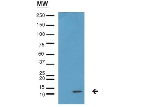
Routinely evaluated by western blot in acid-extracted HeLa cell lysates (Catalog #13-112).

Western Blot Analysis:
0.01-0.1 µg/mL of this lot detected HP1γ protein in acid-extracted HeLa cell lysates (Catalog # 13-112).

Target description

25 kDa

Physical form

Format: Purified
Protein G Purified
Purified mouse monoclonal IgG1 in buffer containing 0.014 M phosphate buffer, pH 7.6, 0.175M NaCl, 0.07% sodium azide, and 30% glycerol. Liquid at -20ºC.

Storage and Stability

Stable for 1 year at -20°C from date of receipt. For maximum recovery of product, centrifuge the vial prior to removing the cap.

Analysis Note

Control
HeLa acid extracts, HEK 293 nuclear extracts.

Other Notes

Concentration: Please refer to the Certificate of Analysis for the lot-specific concentration.
Due to license agreement restrictions, this product cannot be purchased for resale.

Legal Information

UPSTATE is a registered trademark of Merck KGaA, Darmstadt, Germany

Disclaimer

Unless otherwise stated in our catalog or other company documentation accompanying the product(s), our products are intended for research use only and are not to be used for any other purpose, which includes but is not limited to, unauthorized commercial uses, in vitro diagnostic uses, ex vivo or in vivo therapeutic uses or any type of consumption or application to humans or animals.

Not finding the right product?  

Try our Product Selector Tool.

Storage Class

10 - Combustible liquids

wgk_germany

WGK 1


Certificates of Analysis (COA)

Search for Certificates of Analysis (COA) by entering the products Lot/Batch Number. Lot and Batch Numbers can be found on a product’s label following the words ‘Lot’ or ‘Batch’.

Already Own This Product?

Find documentation for the products that you have recently purchased in the Document Library.

Visit the Document Library

A NF-?B-dependent dual promoter-enhancer initiates the lipopolysaccharide-mediated transcriptional activation of the chicken lysozyme in macrophages.
Witham, J; Ouboussad, L; Lefevre, PF
Testing null
Eva Bártová et al.
Differentiation; research in biological diversity, 76(1), 24-32 (2007-06-19)
Human embryonic stem cells (hES) are unique in their pluripotency and capacity for self-renewal. Therefore, we have studied the differences in the level of chromatin condensation in pluripotent and all-trans retinoic acid-differentiated hES cells. Nuclear patterns of the Oct4 (6p21.33)
Rosa M Marion et al.
Cell stem cell, 4(2), 141-154 (2009-02-10)
Telomere shortening is associated with organismal aging. iPS cells have been recently derived from old patients; however, it is not known whether telomere chromatin acquires the same characteristics as in ES cells. We show here that telomeres are elongated in
Thang Van Nguyen et al.
The Journal of experimental medicine, 204(6), 1453-1461 (2007-05-31)
Tumor suppressor p53-dependent apoptosis is critical in suppressing tumorigenesis. Previously, we reported that DNA double-strand breaks (DSBs) at the V(D)J recombination loci induced genomic instability in the developing lymphocytes of nonhomologous end-joining (NHEJ)-deficient, p53-deficient mice, which led to rapid lymphomagenesis.
Marta Kovatcheva et al.
Nature communications, 8(1), 386-386 (2017-09-01)
Senescence is a state of stable cell cycle exit with important implications for development and disease. Here, we demonstrate that the chromatin remodeling enzyme ATRX is required for therapy-induced senescence. ATRX accumulates in nuclear foci and is required for therapy-induced

Our team of scientists has experience in all areas of research including Life Science, Material Science, Chemical Synthesis, Chromatography, Analytical and many others.

Contact Technical Service