
06-1425
Anti-phospho-TAK1 (Ser412) Antibody
from rabbit, purified by affinity chromatography
Synonym(s):
Mitogen-activated protein kinase kinase kinase 7, Transforming growth factor-beta-activated kinase 1, TGF-beta-activated kinase 1
About This Item
Recommended Products

biological source
rabbit
Quality Level
antibody form
affinity isolated antibody
antibody product type
primary antibodies
clone
polyclonal
purified by
affinity chromatography
species reactivity
human, mouse
technique(s)
western blot: suitable
NCBI accession no.
UniProt accession no.
shipped in
wet ice
target post-translational modification
phosphorylation (pSer412)
Gene Information
human ... MAP3K7(6885)
General description
Specificity
Immunogen
Application
Quality
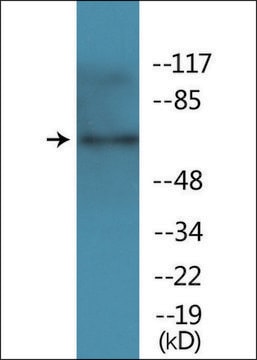
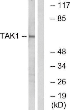
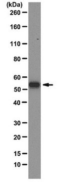
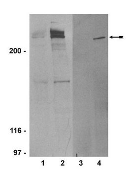
Western Blot Analysis: 1 µg/mL of this antibody detected TAK1 on 10 µg of lambda phosphatase treated and untreated Murine RAW cell lysates.
Target description
Analysis Note
Lambda phosphatase treated and untreated Murine RAW cell lysates
Other Notes
Not finding the right product?
Try our Product Selector Tool.
Storage Class
12 - Non Combustible Liquids
wgk_germany
WGK 1
flash_point_f
Not applicable
flash_point_c
Not applicable
Certificates of Analysis (COA)
Search for Certificates of Analysis (COA) by entering the products Lot/Batch Number. Lot and Batch Numbers can be found on a product’s label following the words ‘Lot’ or ‘Batch’.
Already Own This Product?
Find documentation for the products that you have recently purchased in the Document Library.
Our team of scientists has experience in all areas of research including Life Science, Material Science, Chemical Synthesis, Chromatography, Analytical and many others.
Contact Technical Service