ABN60
Anti-Tryptophan hydroxylase 2 Antibody
from rabbit, purified by affinity chromatography
Sign Into View Organizational & Contract Pricing
All Photos(2)
tryptophan hydroxylase 2, Tryptophan 5-monooxygenase 2, Neuronal tryptophan hydroxylase, tryptophan 5-hydroxylase 2
Recommended Products
biological source
rabbit
Quality Level
antibody form
affinity isolated antibody
antibody product type
primary antibodies
clone
polyclonal
purified by
affinity chromatography
species reactivity
human, rat
species reactivity (predicted by homology)
mouse (immunogen homology)
technique(s)
immunohistochemistry: suitable (paraffin)
western blot: suitable
NCBI accession no.
UniProt accession no.
shipped in
wet ice
target post-translational modification
unmodified
Gene Information
human ... TPH2(121278)
General description
Tryptophan hydroxylase 2 (TPH2) is preferentially expressed in the brain and is thought to play an important role in serotonin synthesis. Recent research has actually associated high levels of TPH2 with risk of suicide. Misfolding of TPH2 may be the cause of a loss of serotonin neuronal function in Parkinson’s disease. Detection and screening of particular mutations in TPH2 has been cited as an approach toward predicting patient sensitivity to potential antidepressant therapies and even the possibility of diagnosis of behavioral disorders.
Immunogen
KLH-conjugated linear peptide corresponding to human Tryptophan hydroxylase 2.
Application
Anti-Tryptophan hydroxylase 2 Antibody detects level of Tryptophan hydroxylase 2 & has been published & validated for use in WB, IH(P).
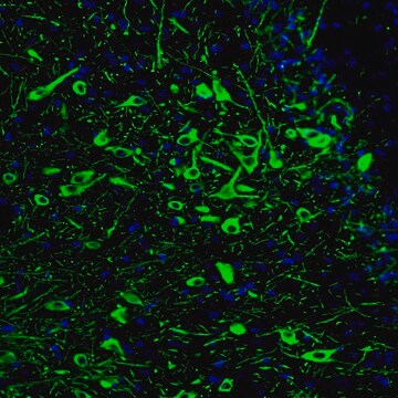
Immunohistochemistry Analysis: 1:400 dilution from a previous lot detected Tryptophan hydroxylase 2 in rat cerebral cortex tissue.
Research Category
Neuroscience
Neuroscience
Research Sub Category
Signaling Neuroscience
Quality
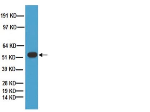
Evaluated by Western Blotting in rat brain tissue lysate.
Western Blotting Analysis: 0.2 µg/mL of this antibody detected Tryptophan hydroxylase 2 on 10 µg of rat brain tissue lysate.
Target description
~56 kDa observed
Physical form
Affinity purified
Purified rabbit polyclonal in buffer containing 0.1 M Tris-Glycine (pH 7.4), 150 mM NaCl with 0.05% sodium azide.
Analysis Note
Control
Rat brain tissue lysate
Disclaimer
Unless otherwise stated in our catalog or other company documentation accompanying the product(s), our products are intended for research use only and are not to be used for any other purpose, which includes but is not limited to, unauthorized commercial uses, in vitro diagnostic uses, ex vivo or in vivo therapeutic uses or any type of consumption or application to humans or animals.
wgk_germany
WGK 1
flash_point_f
Not applicable
flash_point_c
Not applicable
Certificates of Analysis (COA)
Search for Certificates of Analysis (COA) by entering the products Lot/Batch Number. Lot and Batch Numbers can be found on a product’s label following the words ‘Lot’ or ‘Batch’.
Already Own This Product?
Find documentation for the products that you have recently purchased in the Document Library.
Nature communications, 12(1), 3525-3525 (2021-06-12)
Contrasting to the established role of the hypothalamic agouti-related protein (AgRP) neurons in feeding regulation, the neural circuit and signaling mechanisms by which they control energy expenditure remains unclear. Here, we report that energy expenditure is regulated by a subgroup
eLife, 8 (2019-07-30)
Formation of long-range axons occurs over multiple stages of morphological maturation. However, the intrinsic transcriptional mechanisms that temporally control different stages of axon projection development are unknown. Here, we addressed this question by studying the formation of mouse serotonin (5-HT)
Science advances, 7(22) (2021-05-28)
The neural circuitry mechanism that underlies dopaminergic (DA) control of innate feeding behavior is largely uncharacterized. Here, we identified a subpopulation of DA neurons situated in the caudal ventral tegmental area (cVTA) directly innervating DRD1-expressing neurons within the lateral parabrachial
The Journal of neuroscience : the official journal of the Society for Neuroscience, 41(22), 4840-4849 (2021-04-24)
The lateral hypothalamus (LH), together with multiple neuromodulatory systems of the brain, such as the dorsal raphe nucleus (DR), is implicated in arousal, yet interactions between these systems are just beginning to be explored. Using a combination of viral tracing
The Journal of neuroscience : the official journal of the Society for Neuroscience, 37(37), 8863-8875 (2017-08-20)
The ability to predict reward promotes animal survival. Both dopamine neurons in the ventral tegmental area and serotonin neurons in the dorsal raphe nucleus (DRN) participate in reward processing. Although the learning effects on dopamine neurons have been extensively characterized
Our team of scientists has experience in all areas of research including Life Science, Material Science, Chemical Synthesis, Chromatography, Analytical and many others.
Contact Technical Service