Skip to Content
MilliporeSigma
All Photos(1)

Documents

MAB1087

Sigma-Aldrich

Anti-Eosinophil Peroxidase Antibody, clone AHE-1

ascites fluid, clone AHE-1, Chemicon®

Sign Into View Organizational & Contract Pricing

Synonym(s):
Anti-EPO, Anti-EPP, Anti-EPX-PEN, Anti-EPXD
UNSPSC Code:
12352203
eCl@ss:
32160702
NACRES:
NA.41

biological source

mouse

Quality Level

antibody form

ascites fluid

antibody product type

primary antibodies

clone

AHE-1, monoclonal

species reactivity

human

manufacturer/tradename

Chemicon®

technique(s)

immunocytochemistry: suitable

isotype

IgG1

NCBI accession no.

UniProt accession no.

shipped in

dry ice

target post-translational modification

unmodified

Gene Information

human ... EPX(8288)

Specificity

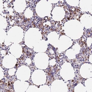
Reacts specifically with human eosinophil peroxidase from eosinophils among peripheral blood cells. Eosinophil peroxidase is felt to play an important role in eosinophil function and perhaps endothelial injury in hypereosinophilic states.

Application

Detect Eosinophil Peroxidase using this Anti-Eosinophil Peroxidase Antibody, clone AHE-1 validated for use in IC.
Final working dilutions must be determined by end user.
Research Category
Inflammation & Immunology
Research Sub Category
Immunoglobulins & Immunology

Physical form

Ascites fluid.

Storage and Stability

Maintain at -20°C in aliquots for up to 12 months. Avoid repeated freeze/thaw cycles.

Legal Information

CHEMICON is a registered trademark of Merck KGaA, Darmstadt, Germany

Disclaimer

Unless otherwise stated in our catalog or other company documentation accompanying the product(s), our products are intended for research use only and are not to be used for any other purpose, which includes but is not limited to, unauthorized commercial uses, in vitro diagnostic uses, ex vivo or in vivo therapeutic uses or any type of consumption or application to humans or animals.

wgk_germany

WGK 1

flash_point_f

Not applicable

flash_point_c

Not applicable


Certificates of Analysis (COA)

Search for Certificates of Analysis (COA) by entering the products Lot/Batch Number. Lot and Batch Numbers can be found on a product’s label following the words ‘Lot’ or ‘Batch’.

Already Own This Product?

Find documentation for the products that you have recently purchased in the Document Library.

Visit the Document Library

Yafeng Song et al.
ISRN inflammation, 2013, 907821-907821 (2013-09-21)
Muscle injury and inflammation (myositis) in a rabbit model of an unilateral muscle overuse were examined. It is unknown if the tachykinin system has a functional role in this situation. In this study, therefore, the neurokinin-1 receptor (NK-1R) expression patterns
Andrea C Bohrer et al.
Cell reports, 40(4), 111144-111144 (2022-07-30)
Influx of eosinophils into the lungs is typically associated with type II responses during allergy and fungal and parasitic infections. However, we previously reported that eosinophils accumulate in lung lesions during type I inflammatory responses to Mycobacterium tuberculosis (Mtb) in
Yafeng Song et al.
BMC musculoskeletal disorders, 14, 134-134 (2013-04-17)
Tachykinins can have pro-inflammatory as well as healing effects during tissue reorganization and inflammation. Recent studies report an up-regulation in the expression of the substance P (SP)-preferred receptor, the neurokinin-1 receptor, in marked muscle inflammation (myositis). There is, however, only
Yafeng Song et al.
PloS one, 7(12), e52230-e52230 (2013-01-04)
It is well established that unilateral exercise can produce contralateral effects. However, it is unclear whether unilateral exercise that leads to muscle injury and inflammation also affects the homologous contralateral muscles. To test the hypothesis that unilateral muscle injury causes
C Spang et al.
Journal of musculoskeletal & neuronal interactions, 17(3), 226-236 (2017-09-02)
The peritendinous connective tissues can have importance in chronic tendon pain. Recently cytokine TNF-α has been suggested to be involved in tendinopathic processes. It is not known how TNF-α and its receptors TNFR1 and TNFR2 are expressed in peritendinous tissues.

Our team of scientists has experience in all areas of research including Life Science, Material Science, Chemical Synthesis, Chromatography, Analytical and many others.

Contact Technical Service