
MAB5316
Anti-Rhodopsin Antibody, clone RET-P1
clone RET-P1, Chemicon®, from mouse
Synonym(s):
Opsin 2
Sign Into View Organizational & Contract Pricing
Select a Size
All Photos(1)
Select a Size
Change View
About This Item
UNSPSC Code:
12352203
eCl@ss:
32160702
NACRES:
NA.41
Recommended Products

biological source
mouse
Quality Level
antibody form
purified immunoglobulin
antibody product type
primary antibodies
clone
RET-P1, monoclonal
species reactivity
human, mouse, rat
manufacturer/tradename
Chemicon®
technique(s)
immunocytochemistry: suitable
western blot: suitable
isotype
IgG1
NCBI accession no.
Specificity
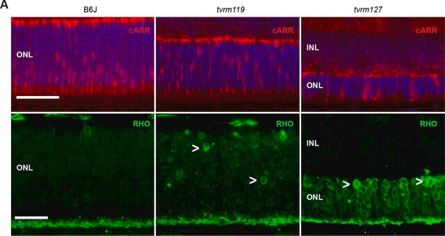
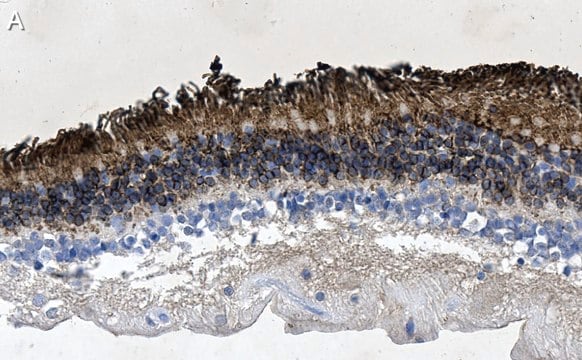
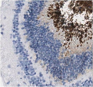
Reacts with a protein of 39 kDa identified as rhodopsin (opsin). MAB5316 specifically labels the axons and synaptic pedicles of the rods.
CELLULAR LOCALIZATION:
Cytoplasmic
CELLULAR LOCALIZATION:
Cytoplasmic
Immunogen
Membrane preparation from adult rat retina.
Application
Detect Rhodopsin using this Anti-Rhodopsin Antibody, clone RET-P1 validated for use in IC & WB.
Immunoblotting: 1 μg/mL
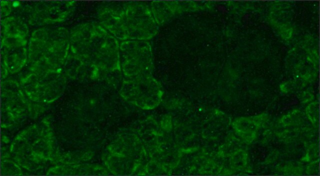
Immunocytochemistry
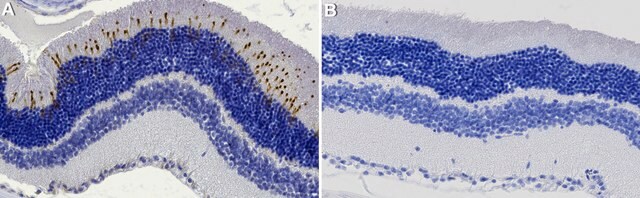
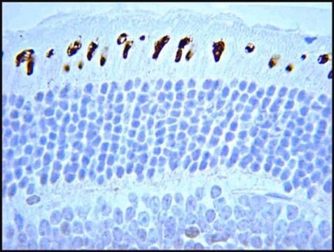
Immunohistochemistry (frozen and formalin/paraffin): 1-2 μg/mL. Staining of formalin fixed tissue sections requires boiling the tissue sections in 10mM citrate buffer, pH 6.0 for 10-20 minutes followed by cooling at room temperature for 20 minutes.
Optimal working dilutions must be determined by end user.
Immunocytochemistry
Immunohistochemistry (frozen and formalin/paraffin): 1-2 μg/mL. Staining of formalin fixed tissue sections requires boiling the tissue sections in 10mM citrate buffer, pH 6.0 for 10-20 minutes followed by cooling at room temperature for 20 minutes.
Optimal working dilutions must be determined by end user.
Research Category
Neuroscience
Neuroscience
Research Sub Category
Sensory & PNS
Sensory & PNS
Physical form
Format: Purified
Purified immunoglobulin. Liquid in 10 mM PBS, pH 7.4 with 0.2% BSA and 15 mM sodium azide.
Storage and Stability
Maintain at 2-8°C in undiluted aliquots for up to 6 months.
Analysis Note
Control
POSITIVE CONTROL:
IMR-5 cells, brain or retina.
POSITIVE CONTROL:
IMR-5 cells, brain or retina.
Other Notes
Concentration: Please refer to the Certificate of Analysis for the lot-specific concentration.
Legal Information
CHEMICON is a registered trademark of Merck KGaA, Darmstadt, Germany
Disclaimer
Unless otherwise stated in our catalog or other company documentation accompanying the product(s), our products are intended for research use only and are not to be used for any other purpose, which includes but is not limited to, unauthorized commercial uses, in vitro diagnostic uses, ex vivo or in vivo therapeutic uses or any type of consumption or application to humans or animals.
Not finding the right product?
Try our Product Selector Tool.
recommended
Product No.
Description
Pricing
Storage Class
12 - Non Combustible Liquids
wgk_germany
WGK 2
flash_point_f
Not applicable
flash_point_c
Not applicable
Certificates of Analysis (COA)
Search for Certificates of Analysis (COA) by entering the products Lot/Batch Number. Lot and Batch Numbers can be found on a product’s label following the words ‘Lot’ or ‘Batch’.
Already Own This Product?
Find documentation for the products that you have recently purchased in the Document Library.
Vibratome sectioning mouse retina to prepare photoreceptor cultures.
Clerin, E; Yang, Y; Forster, V; Fontaine, V; Sahel, JA; Leveillard, T
Journal of Visualized Experiments null
Iman Sahly et al.
The Journal of cell biology, 199(2), 381-399 (2012-10-10)
The mechanisms underlying retinal dystrophy in Usher syndrome type I (USH1) remain unknown because mutant mice lacking any of the USH1 proteins-myosin VIIa, harmonin, cadherin-23, protocadherin-15, sans-do not display retinal degeneration. We found here that, in macaque photoreceptor cells, all
Astrid Zayas-Santiago et al.
Molecular vision, 15, 1-9 (2009-01-13)
Intact adult photoreceptors in culture can be a valuable tool in the search of therapies for retinal degenerations. The major challenge in this technique is that photoreceptors undergo an alteration in cytoarchitecture and loss of outer segment during the cell
Steroids do not prevent photoreceptor degeneration in the light-exposed T4R rhodopsin mutant dog retina irrespective of AP-1 inhibition.
Gu, D; Beltran, WA; Pearce-Kelling, S; Li, Z; Acland, GM; Aguirre, GD
Investigative Ophthalmology & Visual Science null
Canine retina has a primate fovea-like bouquet of cone photoreceptors which is affected by inherited macular degenerations.
Beltran, WA; Cideciyan, AV; Guziewicz, KE; Iwabe, S; Swider, M; Scott, EM; Savina et al.
Testing null
Our team of scientists has experience in all areas of research including Life Science, Material Science, Chemical Synthesis, Chromatography, Analytical and many others.
Contact Technical Service