Skip to Content
MilliporeSigma
All Photos(1)

Key Documents

A3812

Sigma-Aldrich

Anti-Rabbit IgG (whole molecule)–Alkaline Phosphatase antibody produced in goat

affinity isolated antibody, buffered aqueous glycerol solution

Synonym(s):

Anti-Rabbit IgG (whole molecule)–Alkaline Phosphatase antibody produced in goat

Sign Into View Organizational & Contract Pricing


About This Item

MDL number:
UNSPSC Code:
12352203
NACRES:
NA.46

biological source

goat

Quality Level

conjugate

alkaline phosphatase conjugate

antibody form

affinity isolated antibody

antibody product type

secondary antibodies

clone

polyclonal

form

buffered aqueous glycerol solution

species reactivity

rabbit

technique(s)

direct ELISA: 1:30,000
dot blot: 1:30,000
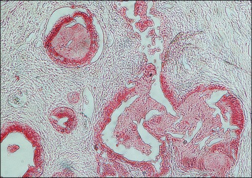
immunohistochemistry (formalin-fixed, paraffin-embedded sections): 1:50
western blot: 1:30,000

shipped in

wet ice

storage temp.

2-8°C

target post-translational modification

unmodified

Looking for similar products? Visit Product Comparison Guide

General description

Anti-rabbit IgG (whole molecule) is produced in goat using purified rabbit IgG as the immunogen. Antibody is isolated from anti-rabbit IgG antiserum by immunospecific purification which removes essentially all goat serum proteins, including immunoglobulins that do not specifically bind to rabbit IgG. The antibody preparation is solid phase adsorbed with human serum proteins to ensure minimal cross reactivity in tissue or cell preparations. Anti-Rabbit IgG is conjugated to alkaline phosphatase by protein cross linking with 0.2% glutaraldehyde.
Specificity of the antiserum is determined by immunoelectrophoresis prior to conjugation, versus normal rabbit serum and rabbit IgG. Identity and purity of the antibody is established by immunoelectrophoresis prior to conjugation. Electrophoresis of the product followed by diffusion versus anti-goat IgG and anti-goat whole serum results in single arcs of precipitation.
IgGs are glycoprotein antibodies that modulate several immune responses. Rabbit IgGs against target proteins are often used as primary antibodies in various research applications. Thus, secondary anti-rabbit IgGs conjugated to a detectable substrate are useful tools for the analysis of target proteins.
Immunoglobulin G (IgG) belongs to the immunoglobulin family and is a widely expressed serum antibody. It consists of a gamma (γ) heavy chain in the constant (C) region. The monomeric 150kDa structure of IgG constitutes two identical heavy chains and two identical light chains with molecular weight of 50kDa and 25kDa, respectively. The primary structure of this antibody also contains disulfide bonds involved in linking the two heavy chains, linking the heavy and light chains and resides inside the chains. IgG is further subdivided into four classes namely, IgG1, IgG2, IgG3, and IgG4 with different heavy chains, named γ1, γ2, γ3, and γ4, respectively. Limited digestion using papain cleaves the antibody into three fragments, two of which are identical and contain the antigen-binding activity (Fab fragments). The third fragment does not possess antigen-binding activity and is known as fragment crystallizable (Fc).

Immunogen

Purified rabbit IgG

Application

Citrullination of antithrombin by PADI4 was analyzed by ELISA using alkaline phosphatase conjugated goat anti-rabbit IgG as the secondary diluted at 1:5000 in 0.05M carbonate/bicarbonate buffer (Ph 9.6).
Goat anti-rabbit IgG (whole molecule)-alkaline phosphatase antibody can be used for western blot and ELISA
It can also be used for immunohistochemistry (1:50) and dot bot (1:30,000) assays.

Physical form

Solution in 0.05 M Tris buffer, pH 8.0, containing 1 mM MgCl2, 10 mM glycine, 1% bovine serum albumin, 50% glycerol and 15 mM sodium azide

Preparation Note

Adsorbed to reduce background staining with human samples.

Disclaimer

Unless otherwise stated in our catalog or other company documentation accompanying the product(s), our products are intended for research use only and are not to be used for any other purpose, which includes but is not limited to, unauthorized commercial uses, in vitro diagnostic uses, ex vivo or in vivo therapeutic uses or any type of consumption or application to humans or animals.

Not finding the right product?  

Try our Product Selector Tool.

Storage Class

10 - Combustible liquids

wgk_germany

WGK 2


Certificates of Analysis (COA)

Search for Certificates of Analysis (COA) by entering the products Lot/Batch Number. Lot and Batch Numbers can be found on a product’s label following the words ‘Lot’ or ‘Batch’.

Already Own This Product?

Find documentation for the products that you have recently purchased in the Document Library.

Visit the Document Library

Ana C Ribeiro et al.
Molecules (Basel, Switzerland), 19(12), 20350-20373 (2014-12-10)
Legume lectins comprise a structurally related, Ca/Mn-dependent, widespread, abundant and well characterized lectin family when compared to the large number of lectins from other sources described in the literature. Strangely enough, no specific function has been assigned to them aside
Focal cerebral ischemia induces increased myelin basic protein and growth-associated protein-43 gene transcription in peri-infarct areas in the rat brain
Gregersen R, et al.
Experimental Brain Research. Experimentelle Hirnforschung. Experimentation Cerebrale, 138(3), 384-392 (2001)
Dorthe B Corlin et al.
Clinical chemistry, 51(7), 1177-1184 (2005-05-14)
Patients on chronic hemodialysis are prone to develop amyloid deposits of misfolded beta(2)-microglobulin (beta(2)M) in osteoarticular tissues. beta(2)M with various deletions/truncations and chemical modifications has been found together with structurally intact beta(2)M in extracts of beta(2)M amyloid fibrils. The state
Helle Hvilsted Nielsen et al.
Journal of neuropathology and experimental neurology, 68(8), 845-856 (2009-07-17)
Acute multiple sclerosis lesions are characterized by accumulation of T cells and macrophages, destruction of myelin and oligodendrocytes, and axonal damage. There is, however, limited information on neuroimmune interactions distal to sites of axonal damage in the T cell-infiltrated central
Hans-Christian Slotved et al.
Journal of clinical microbiology, 45(9), 2929-2936 (2007-07-20)
We identified three isolates of Streptococcus agalactiae (group B streptococcus [GBS]), of human origin, which failed to react with antisera against any of the nine known GBS serotypes. Polyclonal rabbit antisera raised against these isolates and standard GBS typing sera

Our team of scientists has experience in all areas of research including Life Science, Material Science, Chemical Synthesis, Chromatography, Analytical and many others.

Contact Technical Service