Skip to Content
MilliporeSigma
All Photos(2)

Documents

MABN742

Sigma-Aldrich

Anti-Neuroligin-1, clone 1C9.1 Antibody

clone 1C9.1, from mouse

Synonym(s):

neuroligin 1, NLGN1, neuroligin-1

Sign Into View Organizational & Contract Pricing


About This Item

UNSPSC Code:
12352203
eCl@ss:
32160702
NACRES:
NA.41

biological source

mouse

Quality Level

antibody form

purified immunoglobulin

antibody product type

primary antibodies

clone

1C9.1, monoclonal

species reactivity

human, rat

technique(s)

immunohistochemistry: suitable
western blot: suitable

isotype

IgG2aκ

NCBI accession no.

UniProt accession no.

shipped in

wet ice

target post-translational modification

unmodified

Gene Information

human ... NLGN1(22871)

General description

Neuroligin-1 is a neuronal cell surface protein belonging to the type-B carboxylesterase/lipase family. It is a necessary component in the maturation of excitatory synapses for their normal, functional development, and is necessary to the regulation of synaptic plasticity and the development of long-term memory within the adult amygdala in mammals. It is believed to participate in cell-cell-interaction through the assembly of intracellular junction by the binding of beta-neurexins, and may also be a factor in the maintenance and assembly of synaptic junctions. It is also thought to have involvement in excitatory synaptic specification. Within brain tissue, Neuroligin-1 is primarily observed in neurons and spinal cord.

Immunogen

Epitope: Extracellular domain
GST-tagged recombinant protein corresponding to the extracellular domain of Human Neuroligin-1.

Application

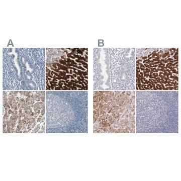
Immunohistochemistry Analysis: A 1:50 dilution from a representative lot detected Neuroligin-1 in human cerebral cortex, rat cerebellum and rat cerebral cortex tissue.
Research Category
Neuroscience

Neuroscience
Research Sub Category
Synapse & Synaptic Biology

Growth Cones & Axon Guidance
This Anti-Neuroligin-1, clone 1C9.1 Antibody is validated for use in Western Blotting and Immunohistochemistry for the detection of Neuroligin-1.

Quality

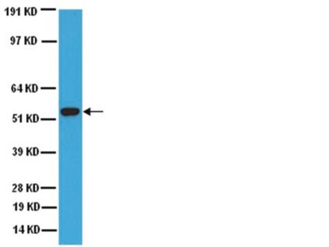
Evaluated by Western Blotting in rat brain tissue lysate.

Western Blotting Analysis: 1.0 µg/mL of this antibody detected Neuroligin-1 in 10 µg of rat brain tissue lysate.

Target description

~94 kDa observed

Physical form

Format: Purified
Protein G Purified
Purified mouse monoclonal IgG2aκ in buffer containing 0.1 M Tris-Glycine (pH 7.4), 150 mM NaCl with 0.05% sodium azide.

Storage and Stability

Stable for 1 year at 2-8°C from date of receipt.

Other Notes

Concentration: Please refer to lot specific datasheet.

Disclaimer

Unless otherwise stated in our catalog or other company documentation accompanying the product(s), our products are intended for research use only and are not to be used for any other purpose, which includes but is not limited to, unauthorized commercial uses, in vitro diagnostic uses, ex vivo or in vivo therapeutic uses or any type of consumption or application to humans or animals.

Not finding the right product?  

Try our Product Selector Tool.

Storage Class

12 - Non Combustible Liquids

wgk_germany

WGK 1

flash_point_f

Not applicable

flash_point_c

Not applicable


Certificates of Analysis (COA)

Search for Certificates of Analysis (COA) by entering the products Lot/Batch Number. Lot and Batch Numbers can be found on a product’s label following the words ‘Lot’ or ‘Batch’.

Already Own This Product?

Find documentation for the products that you have recently purchased in the Document Library.

Visit the Document Library

Sebnem Ece Eksi et al.
Nature communications, 12(1), 7292-7292 (2021-12-17)
Identifying precise molecular subtypes attributable to specific stages of localized prostate cancer has proven difficult due to high levels of heterogeneity. Bulk assays represent a population-average, which mask the heterogeneity that exists at the single-cell level. In this work, we
Julien Dufort-Gervais et al.
Scientific reports, 10(1), 6956-6956 (2020-04-26)
Synapse loss occurs early and correlates with cognitive decline in Alzheimer's disease (AD). Synaptotoxicity is driven, at least in part, by amyloid-beta oligomers (Aβo), but the exact synaptic components targeted by Aβo remain to be identified. We here tested the

Our team of scientists has experience in all areas of research including Life Science, Material Science, Chemical Synthesis, Chromatography, Analytical and many others.

Contact Technical Service