MABT886
Anti-VE-Cadherin Antibody, clone Vli37
culture supernatant, clone Vli37, from rabbit
Sign Into View Organizational & Contract Pricing
All Photos(5)
Cadherin-5, CD144, Vascular endothelial cadherin, VE-Cad, VE-cadherin, VEcad
Recommended Products
biological source
rabbit
Quality Level
antibody form
culture supernatant
antibody product type
primary antibodies
clone
Vli37, monoclonal
species reactivity
bovine, mouse
species reactivity (predicted by homology)
rat (based on 100% sequence homology)
technique(s)
immunocytochemistry: suitable
immunohistochemistry: suitable (paraffin)
western blot: suitable
isotype
IgG
NCBI accession no.
UniProt accession no.
shipped in
dry ice
target post-translational modification
unmodified
Gene Information
mouse ... Cdh5(12562)
General description
Cadherin-5 (UniProt P55284; also known as Vascular endothelial cadherin, VE-Cad, VE-cadherin, VEcad, CD144) is encoded by the Cdh5 gene (Gene ID 12562) in murine species. Cadherins (calcium-dependent adhesion) are type-1 transmembrane proteins that form adherens junctions in tissues and play important roles in mediating cell adhesion. Cadherins are designated with a prefix that specifies their tissue association. Vascular endothelial-cadherin (VE-cadherin) is the transmembrane component of the endothelial adherens junction between vascular endothelial cells (ECs) and plays a pivotal role in endothelium integrity and in the control of vascular permeability. One characteristic of VEGF-induced vascular permeability is the phosphorylation of VE-cadherin, which leads to VE-cadherin internalization and the destabilization of adherens junctions. Likewise, VE-cadherin overexpression is shown to decrease the permeability of endothelial monolayers in vitro. VE-cadherin is initially produced with a signal peptide (a.a. 1-24) and a propeptide (a.a. 25-45) sequence, the removal of which yields tthe mature protein containing a large extracellular region (a.a. 46-599) with five cadherine repeats (a.a. 46-149, 150-256, 257-371, 372-476, 477-593), followed by a transmembrane segment (a.a. 600-620) and a cytoplasmic domain (a.a. 621-784).
Specificity
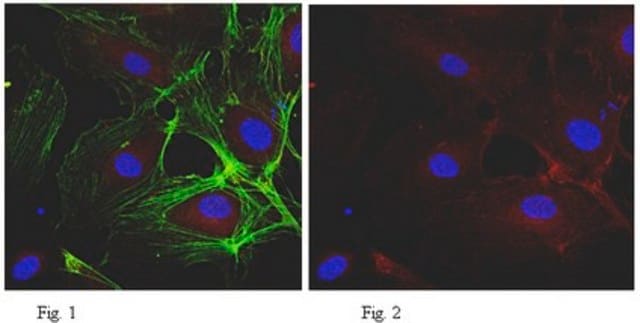
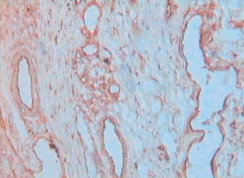
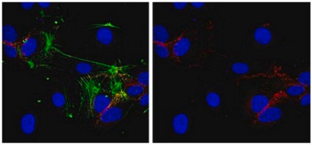
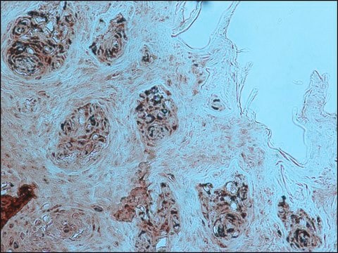
Clone Vli37 stains endothelial adherens junctions by immunocytochemistry and immunohistochemistry.
Immunogen
Epitope: Near C-terminus.
Linear peptide corresponding to the C-terminal cytoplasmic domain sequence of mouse VE-Cadherin.
Application
Research Category
Cell Structure
Cell Structure
Research Sub Category
Adhesion (CAMs)
Adhesion (CAMs)
This Anti-VE-Cadherin Antibody, clone Vli37 is validated for use in Western Blotting, Immunohistochemistry (Paraffin), Immunocytochemistry for the detection of VE-Cadherin.
Western Blotting Analysis: An 1:500 dilution of this antibody detected VE-Cadherin in 10 µg of mouse brain endothelial bEnd.3 cell lysate.
Western Blotting Analysis: An 1:2,000 dilution from a representative lot detected VE-cadherin expression in lysastes from mouse lung tissue, mouse brain endothelial bEnd.3 cells, and bovine aortic endothelial GM7372 cells (Courtesy of Dr. Volkhard Lindner, Maine Medical Center Research Institute, Scarborough, ME).
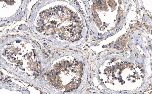
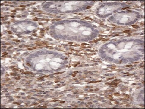
Immunohistochemistry Analysis: An 1:250-5,000 dilution from a representative lot detected VE-cadherin immunoreactivity in formalin-fixed, paraffin-embeded mouse lung, aorta, and liver tissue sections (Courtesy of Dr. Volkhard Lindner, Maine Medical Center Research Institute, Scarborough, ME).
Immunocytochemistry Analysis: An 1:500-5,000 dilution from a representative lot detected VE-cadherin immunoreactivity in bovine aortic endothelial GM7372 cells (Courtesy of Dr. Volkhard Lindner, Maine Medical Center Research Institute, Scarborough, ME).
Quality
Evaluated by Western Blotting in mouse lung tissue lysate.
Western Blotting Analysis: An 1:500 dilution of this antibody detected VE-Cadherin in 10 µg of mouse lung tissue lysate.
Target description
~110 observed. Target band size appears larger than the calculated molecular weight of 83.05 kDa due to glycosylation.
Physical form
Rabbit monoclonal IgG in supernatant with 0.05% sodium azide.
Unpurified
Storage and Stability
Stable for 1 year at -20°C from date of receipt.
Handling Recommendations: Upon receipt and prior to removing the cap, centrifuge the vial and gently mix the solution. Aliquot into microcentrifuge tubes and store at -20°C. Avoid repeated freeze/thaw cycles, which may damage IgG and affect product performance.
Other Notes
Concentration: Please refer to lot specific datasheet.
Disclaimer
Unless otherwise stated in our catalog or other company documentation accompanying the product(s), our products are intended for research use only and are not to be used for any other purpose, which includes but is not limited to, unauthorized commercial uses, in vitro diagnostic uses, ex vivo or in vivo therapeutic uses or any type of consumption or application to humans or animals.
wgk_germany
WGK 2
Certificates of Analysis (COA)
Search for Certificates of Analysis (COA) by entering the products Lot/Batch Number. Lot and Batch Numbers can be found on a product’s label following the words ‘Lot’ or ‘Batch’.
Already Own This Product?
Find documentation for the products that you have recently purchased in the Document Library.
Journal of vascular research, 61(2), 68-76 (2024-01-15)
While multiple factors influence coronary artery bypass graft (CABG) success rates, preserving saphenous vein endothelium during surgery may improve patency. Standard preparations include saphenous vein preparation in heparinized saline (saline) which can result in endothelial loss and damage. Here, we
Arteriosclerosis, thrombosis, and vascular biology, 38(11), 2691-2705 (2018-10-26)
Objective- Blood-CNS (central nervous system) barrier defects are implicated in retinopathies, neurodegenerative diseases, stroke, and epilepsy, yet, the pathological mechanisms downstream of barrier defects remain incompletely understood. Blood-retina barrier (BRB) formation and retinal angiogenesis require β-catenin signaling induced by the
Cell and tissue research, 395(1), 81-103 (2023-11-30)
Endothelial cells of mammalian blood vessels have multiple levels of heterogeneity along the vascular tree and among different organs. Further heterogeneity results from blood flow turbulence and variations in shear stress. In the aorta, vascular endothelial protein tyrosine phosphatase (VE-PTP)
EMBO molecular medicine, 15(4), e16128-e16128 (2023-02-07)
Vascular endothelial protein tyrosine phosphatase (VE-PTP) influences endothelial barrier function by regulating the activation of tyrosine kinase receptor Tie2. We determined whether this action is linked to the development of atherosclerosis by examining the influence of arterial shear stress on
Our team of scientists has experience in all areas of research including Life Science, Material Science, Chemical Synthesis, Chromatography, Analytical and many others.
Contact Technical Service