Skip to Content
MilliporeSigma
All Photos(1)

Key Documents

291M-1

Sigma-Aldrich

MUC2 (MRQ-18) Mouse Monoclonal Antibody

Sign Into View Organizational & Contract Pricing


About This Item

UNSPSC Code:
12352203
NACRES:
NA.41

biological source

mouse

Quality Level

100
500

conjugate

unconjugated

antibody form

culture supernatant

antibody product type

primary antibodies

clone

MRQ-18, monoclonal

description

For In Vitro Diagnostic Use in Select Regions (See Chart)

form

buffered aqueous solution

species reactivity

human

packaging

vial of 0.1 mL concentrate (291M-14)
vial of 0.5 mL concentrate (291M-15)
bottle of 1.0 mL predilute (291M-17)
vial of 1.0 mL concentrate (291M-16)
bottle of 7.0 mL predilute (291M-18)

manufacturer/tradename

Cell Marque

technique(s)

immunohistochemistry (formalin-fixed, paraffin-embedded sections): 1:50-1:200

isotype

IgG1κ

control

colon

shipped in

wet ice

storage temp.

2-8°C

visualization

cytoplasmic

Gene Information

human ... MUC2(4583)

General description

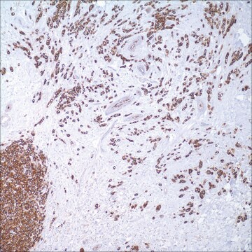
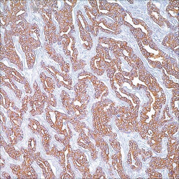
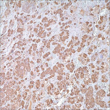
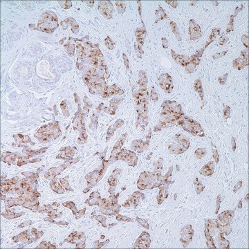
Mucins are a diverse group of complex, highly glycosylated extracellular proteins. Mucin 2 (MUC2) protects the gastric and intestinal epithelium from chemical and mechanical injury. Anti-MUC2 reactivity is normally seen in goblet cells of the small intestine and colon and is associated with mucinous carcinomas, including those of the gastrointestinal tract and ovary. MUC2 immunohistochemistry is useful for identifying colonic, gastric, and esophageal carcinomas.
Mucins are high molecular weight glycoproteins which constitute the major component of the mucus layer that protects the gastric epithelium from chemical and mechanical aggressions. In humans, at least 14 mucin genes have been identified that code for the mucin proteins. They are designated as MUC1, MUC2, MUC3, MUC4, MUC5AC, MUC5B , MUC6 , MUC7, MUC8, MUC9, MUC11, MUC12, MUC13 and MUC16. Mucins share a common feature of a tandem-repeat domain rich in serine and threonine residues. These amino acid residues are potential O-glycosylation sites for attachment of the O-glycan chains that constitute up to 80% of the molecular weight of the final mucin glycoprotein.

The heterogeneous pattern of mucin expression, including the expression of the intestinal mucin MUC2, may provide new insights into the differentiation pathways of gastric carcinoma. The pattern of mucin expression may also be used as a clue to bring new insights into the biological behaviour of distinct clinicopathological entities related to the localisation of gastric carcinoma, namely proximal and distal gastric carcinomas. Pinto-de-Sousa et al. have shown in a comprehensive study of gastric carcinomas evaluated for expression of several mucins (MUC1, MUC2, MUC5AC and MUC6) that: (1) mucin expression is associated with tumour type (MUC5AC with diffuse and infiltrative carcinomas and MUC2 with mucinous carcinomas) but not with the clinico-biological behaviour of the tumours; and (2) mucin expression is associated with tumour location (MUC5AC with antrum carcinomas and MUC2 with cardia carcinomas), indirectly reflecting differences in tumour differentiation according to tumour location.

The following generalities apply to the patterns of Mucin expression:

MUC1 expression: apical surfaces of most epithelial cells in breast, GI, respiratory, and GU tracts.

MUC2 expression: specifically expressed in goblet cells of the small intestine & colon.

Colonic CAs – 65%, Gastric CAs – 42%, Esophageal CAs – 17%

Rare outside of GI tract – with exception of; mucinous ca of breast, clear cell-type CAs of the ovary.

Quality


IVD

IVD

IVD

RUO

Linkage

MUC2 Positive Control Slides, Product No. 291S, are available for immunohistochemistry (formalin-fixed, paraffin-embedded sections).

Physical form

Solution in Tris Buffer, pH 7.3-7.7, with 1% BSA and <0.1% Sodium Azide

Preparation Note

Download the IFU specific to your product lot and formatNote: This requires a keycode which can be found on your packaging or product label.

Other Notes

For Technical Service please contact: 800-665-7284 or email: service@cellmarque.com

Legal Information

Cell Marque is a trademark of Merck KGaA, Darmstadt, Germany

Not finding the right product?  

Try our Product Selector Tool.


Choose from one of the most recent versions:

Certificates of Analysis (COA)

Lot/Batch Number

Don't see the Right Version?

If you require a particular version, you can look up a specific certificate by the Lot or Batch number.

Already Own This Product?

Find documentation for the products that you have recently purchased in the Document Library.

Visit the Document Library

Seog-Yun Park et al.
Archives of pathology & laboratory medicine, 131(10), 1561-1567 (2007-10-10)
Although identification of the primary tumor in patients with metastatic adenocarcinoma has a profound clinical impact, diagnosing the organ of origin is frequently difficult. Because none of the individual immunohistochemical markers used for tissue identification are both site specific and
Emad A Rakha et al.
Modern pathology : an official journal of the United States and Canadian Academy of Pathology, Inc, 18(10), 1295-1304 (2005-06-25)
Mucins are a large family of glycoproteins expressed by many epithelial cells and their malignant counterparts. Much interest has been focused on expression of its members in breast cancer because of their potential role as prognostic indicators and their involvement
T Mizoshita et al.
Histology and histopathology, 22(3), 251-260 (2006-12-14)
We have previously demonstrated links between clinicopathological findings and phenotypes using several gastric and intestinal phenotypic markers in stomach and pancreatic cancers. However, the clinicopathological significance of the phenotype and Cdx2 expression has hitherto remained unclear in colorectal carcinogenesis. We
Emmanuelle Leteurtre et al.
World journal of gastroenterology, 12(21), 3324-3331 (2006-05-31)
To investigate the expression of the four secreted gel-forming mucins (MUC2, MUC5AC, MUC5B and MUC6) in a series of gastric carcinomas, classified according Lauren's, Mulligan's, WHO and Goseki's classifications, with special attention to all the different components (major and minor)
P Chaves et al.
Diseases of the esophagus : official journal of the International Society for Diseases of the Esophagus, 18(6), 383-387 (2005-12-13)
Intestinal metaplasia is a prerequisite criterion for the diagnosis of Barrett's metaplasia and the sole columnar esophageal lining associated with malignancy. It is recognized by the presence of goblet cells, but columnar non-goblet elements, producing gastric or intestinal proteins, are

Our team of scientists has experience in all areas of research including Life Science, Material Science, Chemical Synthesis, Chromatography, Analytical and many others.

Contact Technical Service