Skip to Content
MilliporeSigma
All Photos(1)

Documents

402M-1

Sigma-Aldrich

Cathepsin K (3F9) Mouse Monoclonal Antibody

Sign Into View Organizational & Contract Pricing


About This Item

UNSPSC Code:
12352203
NACRES:
NA.41

biological source

mouse

Quality Level

100
500

conjugate

unconjugated

antibody form

culture supernatant

antibody product type

primary antibodies

clone

3F9, monoclonal

description

For In Vitro Diagnostic Use in Select Regions (See Chart)

form

buffered aqueous solution

species reactivity

human

packaging

vial of 0.1 mL concentrate (402M-14)
vial of 0.5 mL concentrate (402M-15)
bottle of 1.0 mL predilute (402M-17)
vial of 1.0 mL concentrate (402M-16)
bottle of 7.0 mL predilute (402M-18)

manufacturer/tradename

Cell Marque

technique(s)

immunohistochemistry: 1:25-1:100

isotype

IgG2b

shipped in

wet ice

storage temp.

2-8°C

Gene Information

human ... CTSK(1513)

General description

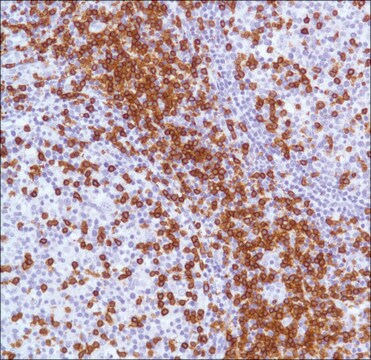
Cathepsin K is a lysosomal papain–like cystine proteinase and can be expressed in normal osteoblasts, fibroblasts, and skin.1 Recently, cathepsin K has been reported as useful in distinguishing translocation renal cell carcinoma (RCC) from other subtypes of RCC.1 Mortignoni et al. tested 17 cases of translocation RCC and found that cathepsin K stained 6 of 8 cases of translocation RCC with t(X;1), all seven cases of translocation RCC with t(6;11), but was negative for one case with t(X;3) and one with t(X;17).1 Additionally, the study compared IHC detection of TFE3 antibody in the 17 cases and demonstrated that all translocation RCC with t(6;11) were completely negative for TFE3, but positive for cathepsin K; all other translocation RCC with different chromosomal tranlocations other than t(X;1) expressed TFE3.1 Thus, it is recommended that antibodies against both TFE3 and cathepsin K should be used for evaluating translocation renal cell carcinomas.1 None of the 305 other renal cell neoplasms representing the most common renal cell neoplasm subtypes were positive for cathepsin K.1 Zheng et al. reported only 2.7% of 1,140 carcinomas from various organs exhibited cathepsin K labeling, indicating that among carcinomas cathepsin K labeling is highly specific for translocation RCC and can be used to help differentiate translocation RCC from other carcinomas.2

Quality


IVD

IVD

IVD

RUO

Physical form

Solution in Tris Buffer, pH 7.3-7.7, with 1% BSA and <0.1% Sodium Azide

Preparation Note

Download the IFU specific to your product lot and formatNote: This requires a keycode which can be found on your packaging or product label.

Other Notes

For Technical Service please contact: 800-665-7284 or email: service@cellmarque.com

Legal Information

Cell Marque is a trademark of Merck KGaA, Darmstadt, Germany

Not finding the right product?  

Try our Product Selector Tool.

Storage Class

12 - Non Combustible Liquids

wgk_germany

WGK 2

flash_point_f

Not applicable

flash_point_c

Not applicable


Certificates of Analysis (COA)

Search for Certificates of Analysis (COA) by entering the products Lot/Batch Number. Lot and Batch Numbers can be found on a product’s label following the words ‘Lot’ or ‘Batch’.

Already Own This Product?

Find documentation for the products that you have recently purchased in the Document Library.

Visit the Document Library

G Martignoni et al.
Modern pathology : an official journal of the United States and Canadian Academy of Pathology, Inc, 22(8), 1016-1022 (2009-04-28)
The microphthalmia transcription factor/transcription factor E (TFE)-family translocation renal cell carcinomas bear specific translocations that result in overexpression of TFE3 or TFEB. TFE3 fusion gene product overexpression occurs as consequence of different translocations involving chromosome Xp11.2, whereas TFEB overexpression is
Gang Zheng et al.
American journal of clinical pathology, 139(2), 151-159 (2013-01-29)
Cathepsin K is consistently and diffusely expressed in alveolar soft part sarcoma (ASPS) and a subset of translocation renal cell carcinomas (RCCs). However, cathepsin K expression in human neoplasms has not been systematically analyzed. We constructed tissue microarrays (TMA) from

Our team of scientists has experience in all areas of research including Life Science, Material Science, Chemical Synthesis, Chromatography, Analytical and many others.

Contact Technical Service