Skip to Content
MilliporeSigma
All Photos(2)

Key Documents

C5585

Sigma-Aldrich

Monoclonal Anti-Biotin–Cy3 antibody produced in mouse

clone BN-34, purified immunoglobulin, buffered aqueous solution

Synonym(s):

Monoclonal Anti-Biotin

Sign Into View Organizational & Contract Pricing


About This Item

UNSPSC Code:
12352203
NACRES:
NA.46

biological source

mouse

Quality Level

conjugate

CY3 conjugate

antibody form

purified immunoglobulin

antibody product type

primary antibodies

clone

BN-34, monoclonal

form

buffered aqueous solution

technique(s)

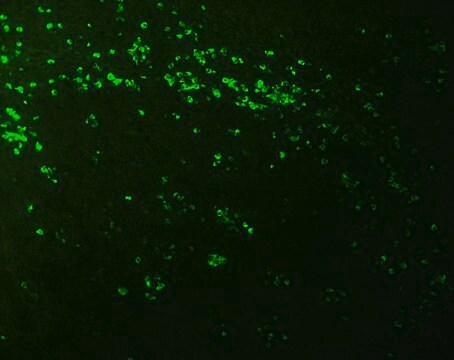
immunohistochemistry (formalin-fixed, paraffin-embedded sections): 1:400
indirect immunofluorescence: 1:200

isotype

IgG1

shipped in

wet ice

storage temp.

2-8°C

target post-translational modification

unmodified

Looking for similar products? Visit Product Comparison Guide

General description

Monoclonal Anti-Biotin (mouse IgG1 isotype) is derived from the BN-34 hybridoma produced by the fusion of mouse myeloma cells and splenocytes from an immunized mouse. Biotin is a naturally occurring vitamin and co-enzyme required by cells in living organisms or in culture.
The antibody recognizes free biotin and biocytin using competitive ELISA. Using indirect ELISA or immuno­histo­chemical staining, the antibody also recognizes biotin conjugated to various immunoglobulins. Specificity is evaluated using biotinylated goat antibodies to human and rabbit antigens coated on multiwell plates.

Immunogen

Biotin-KLH conjugate

Application

Monoclonal Anti-Biotin−Cy3 antibody produced in mouse has been used in:
  • fluorescent microscopy
  • RNA fluorescence in situ hybridization (FISH){36
  • immunostaining

The antibody may be used in amplification techniques, ELISA, immunoblotting, in situ nucleic acid hybridization, flow cytometry, image analysis or confocal microscopy.
In some applications, localization of biotinylated probes with avidin produces high background levels. Anti-biotin reagents may be substituted for avidin to decrease non-specific binding.

Biochem/physiol Actions

Cy dyes are pH insensitive fluorophores that are used to label, DNA, RNA and proteins in a variety of biological assays.
Monoclonal Anti-Biotin- Cy3 can be used in many other applications where biotin can be introduced as target label. It can be used in detection of DNA and mRNA by sensitive non-isotopic in situ hybridization. It may also be used for detection of microinjected biotin-haptenized cytoskeletal proteins allowing examination of the pattern of incorporation and turnover of cytoskeletal proteins in living cells.

Physical form

Solution in phosphate buffered saline, pH 7.4, containing 1% bovine serum albumin and 15 mM sodium azide.

Legal Information

Cy is distributed under license from Amersham Biosciences Limited.
Cy3 is a trademark of Cytiva

Disclaimer

Unless otherwise stated in our catalog or other company documentation accompanying the product(s), our products are intended for research use only and are not to be used for any other purpose, which includes but is not limited to, unauthorized commercial uses, in vitro diagnostic uses, ex vivo or in vivo therapeutic uses or any type of consumption or application to humans or animals.

Not finding the right product?  

Try our Product Selector Tool.

Storage Class

10 - Combustible liquids

wgk_germany

nwg

flash_point_f

Not applicable

flash_point_c

Not applicable

ppe

Eyeshields, Gloves, multi-purpose combination respirator cartridge (US)


Certificates of Analysis (COA)

Search for Certificates of Analysis (COA) by entering the products Lot/Batch Number. Lot and Batch Numbers can be found on a product’s label following the words ‘Lot’ or ‘Batch’.

Already Own This Product?

Find documentation for the products that you have recently purchased in the Document Library.

Visit the Document Library

Jennifer A Ralph et al.
The American journal of pathology, 170(3), 1121-1133 (2007-02-27)
Peripheral corticotropin-releasing hormone (CRH) is an important regulator of localized inflammatory responses. The aim of this study is to define the pathological signaling pathways in which peripheral CRH receptor-mediated responses reside. We report that PECAM-1-expressing synovial membrane endothelial cells are
Cyril Charbonnel et al.
Plant physiology, 177(1), 311-327 (2018-04-07)
Despite intensive searches, few proteins involved in telomere homeostasis have been identified in plants. Here, we used pull-down assays to identify potential telomeric interactors in the model plant species Arabidopsis (Arabidopsis thaliana). We identified the candidate protein GH1-HMGA1 (also known
High-mobility group nucleosome-binding domain 2 protein inhibits the invasion of Klebsiella pneumoniae into mouse lungs in vivo
Zheng S, et al.
Molecular Medicine Reports, 12(1), 1279-1285 (2015)
Qun Fang et al.
Medical science monitor : international medical journal of experimental and clinical research, 22, 3506-3513 (2016-10-04)
BACKGROUND In this study, we aimed to investigate the association between UCA1 and miR-27b in gastric cancer and further study their involvement in multi-drug resistance (MDR) of gastric cancer. MATERIAL AND METHODS The microarray data of dysregulated lncRNAs in gastric
Surface plasmon excitation in semitransparent inverted polymer photovoltaic devices and their applications as label-free optical sensors
Park B, et al.
Light: Science & Applications, 3(12), e222-e222 (2014)

Our team of scientists has experience in all areas of research including Life Science, Material Science, Chemical Synthesis, Chromatography, Analytical and many others.

Contact Technical Service