Skip to Content
MilliporeSigma
All Photos(6)

Key Documents

HPA005533

Sigma-Aldrich

Anti-MEF2C antibody produced in rabbit

enhanced validation

Prestige Antibodies® Powered by Atlas Antibodies, affinity isolated antibody, buffered aqueous glycerol solution

Synonym(s):

Anti-Myocyte-specific enhancer factor 2C antibody produced in rabbit

Sign Into View Organizational & Contract Pricing


About This Item

UNSPSC Code:
12352203
Human Protein Atlas Number:
NACRES:
NA.43

biological source

rabbit

Quality Level

conjugate

unconjugated

antibody form

affinity isolated antibody

antibody product type

primary antibodies

clone

polyclonal

product line

Prestige Antibodies® Powered by Atlas Antibodies

form

buffered aqueous glycerol solution

species reactivity

human

enhanced validation

orthogonal RNAseq
recombinant expression
Learn more about Antibody Enhanced Validation

technique(s)

immunofluorescence: 0.25-2 μg/mL
immunohistochemistry: 1:50-1:200
western blot: 0.04-0.4 μg/mL

immunogen sequence

PPNFEMPVSIPVSSHNSLVYSNPVSSLGNPNLLPLAHPSLQRNSMSPGVTHRPPSAGNTGGLMGGDLTSGAGTSAGNGYGNPRNSPGLLVSPGNLNKNMQAKSPPPMNLGMNNRKPDLRVLIPPGSKNTMPSVNQRINN

UniProt accession no.

shipped in

wet ice

storage temp.

−20°C

target post-translational modification

unmodified

Gene Information

human ... MEF2C(4208)

General description

MEF2C (myocyte enhancer factor 2C) belongs to Myocyte enhancer factor 2 (MEF2) protein family, which in turn belongs to a family of transcriptional regulators called MADS (MCMI, agamous, deficiens, serum response factor)-box. Four different forms of MEF2 protein are found in vertebrates, namely, MEF2A, MEF2B, MEF2C, and MEF2D. MEF2C is predominantly expressed in brain and skeletal muscle. It shares the common DNA-binding and dimerization domain present at the N-terminal, with other MEF2 proteins. Alternative splicing produces two MEF2C variants, which lack α exon. These MEF2Cα- are ubiquitously expressed, but at a lower level than MEF2Cα+ variants. MEF2Cα- variants are also less expressed in other tissues, as opposed to brain and heart. Alternative splicing also produces either MEF2Cγ+ or MEF2Cγ- isoforms. MEF2Cγ- is the major isoform expressed in differentiating myocytes and adult tissues. MEF2C is mapped to chromosome 5q14.3.

Immunogen

Myocyte-specific enhancer factor 2C recombinant protein epitope signature tag (PrEST)

Application

Anti-MEF2C antibody is suitable for chromatin immunoprecipitation (ChIP).
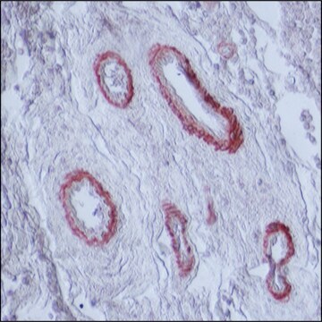
Anti-MEF2C antibody produced in rabbit, a Prestige Antibody, is developed and validated by the Human Protein Atlas (HPA) project . Each antibody is tested by immunohistochemistry against hundreds of normal and disease tissues. These images can be viewed on the Human Protein Atlas (HPA) site by clicking on the Image Gallery link. The antibodies are also tested using immunofluorescence and western blotting. To view these protocols and other useful information about Prestige Antibodies and the HPA, visit sigma.com/prestige.
Applications in which this antibody has been used successfully, and the associated peer-reviewed papers, are given below.
Immunohistochemistry (1 paper)

Biochem/physiol Actions

MEF2C (myocyte enhancer factor 2C) belongs to the family of transcription factors, which regulate gene expression in myocytes, neurons and lymphocytes. BMK1 phosphorylates and activates MEF2C. Serum also induces BMK1-induced phosphorylation of MEF2C, and thus, MEF2C plays a role in serum-dependent early gene expression via BMK1 pathway. Lipopolysaccharides produced during microbial infection activate this protein via phosphorylation by p38. This induces c-jun transcription, which plays a role in inflammation. MEF2C plays a part in neuronal differentiation, as it is expressed in cortical plate, during early development. It is also expressed in neurons having a preference to mature cerebrocortex layers II, IV and VI. MEF2C contributes to early pathogenesis of Parkinson′s disease, as the disruption of MEF2C- PGC1α pathway, leads to neuronal apoptosis due to mitochondrial dysfunction. It is a part of Wnt pathway, and hence plays a role in control of bone mass and turnover.

Features and Benefits

Prestige Antibodies® are highly characterized and extensively validated antibodies with the added benefit of all available characterization data for each target being accessible via the Human Protein Atlas portal linked just below the product name at the top of this page. The uniqueness and low cross-reactivity of the Prestige Antibodies® to other proteins are due to a thorough selection of antigen regions, affinity purification, and stringent selection. Prestige antigen controls are available for every corresponding Prestige Antibody and can be found in the linkage section.

Every Prestige Antibody is tested in the following ways:
  • IHC tissue array of 44 normal human tissues and 20 of the most common cancer type tissues.
  • Protein array of 364 human recombinant protein fragments.

Linkage

Corresponding Antigen APREST79903

Physical form

Solution in phosphate-buffered saline, pH 7.2, containing 40% glycerol and 0.02% sodium azide

Legal Information

Prestige Antibodies is a registered trademark of Merck KGaA, Darmstadt, Germany

Disclaimer

Unless otherwise stated in our catalog or other company documentation accompanying the product(s), our products are intended for research use only and are not to be used for any other purpose, which includes but is not limited to, unauthorized commercial uses, in vitro diagnostic uses, ex vivo or in vivo therapeutic uses or any type of consumption or application to humans or animals.

Not finding the right product?  

Try our Product Selector Tool.

Storage Class

10 - Combustible liquids

wgk_germany

WGK 1

flash_point_f

Not applicable

flash_point_c

Not applicable

ppe

Eyeshields, Gloves, multi-purpose combination respirator cartridge (US)


Certificates of Analysis (COA)

Search for Certificates of Analysis (COA) by entering the products Lot/Batch Number. Lot and Batch Numbers can be found on a product’s label following the words ‘Lot’ or ‘Batch’.

Already Own This Product?

Find documentation for the products that you have recently purchased in the Document Library.

Visit the Document Library

MicroRNA-21 dysregulates the expression of MEF2C in neurons in monkey and human SIV/HIV neurological disease.
Yelamanchili, S.V., et al.
Cell Death and Differentiation, e77, e77-e77 (2010)
Christopher D Clark et al.
Mechanisms of development, 162, 103615-103615 (2020-05-26)
The cardiac homeobox transcription factor Nkx2-5 is a major determinant of cardiac identity and cardiac morphogenesis. Nkx2-5 operates as part of a complex and mutually reinforcing network of early transcription factors of the homeobox, GATA zinc finger and MADS domain
Vittoria Infantino et al.
Gene, 531(2), 355-362 (2013-09-07)
Myocyte enhancer factor 2C (MEF2C) belongs to the MEF2 transcription factors. All products of MEF2 genes have a common amino-terminal DNA binding and dimerization domain. All four vertebrate MEF2 gene transcripts are also alternatively spliced. In the present study we
Dana Trompet et al.
JCI insight, 9(6) (2024-03-22)
Recently, skeletal stem cells were shown to be present in the epiphyseal growth plate (epiphyseal skeletal stem cells, epSSCs), but their function in connection with linear bone growth remains unknown. Here, we explore the possibility that modulating the number of
Scott D Ryan et al.
Cell, 155(6), 1351-1364 (2013-12-03)
Parkinson's disease (PD) is characterized by loss of A9 dopaminergic (DA) neurons in the substantia nigra pars compacta (SNpc). An association has been reported between PD and exposure to mitochondrial toxins, including environmental pesticides paraquat, maneb, and rotenone. Here, using

Our team of scientists has experience in all areas of research including Life Science, Material Science, Chemical Synthesis, Chromatography, Analytical and many others.

Contact Technical Service