Skip to Content
MilliporeSigma
All Photos(1)

Key Documents

SAB4200176

Sigma-Aldrich

Anti-JNK antibody, Mouse monoclonal

clone 1C2, purified from hybridoma cell culture

Synonym(s):

Anti-JNK-46, Anti-JNK1, Anti-JNK1A2, Anti-JNK21B1/2, Anti-JUN N-terminal kinase, Anti-MAP kinase 8, Anti-MAPK8, Anti-PRKM8, Anti-SAPK1, Anti-stress-activated protein kinase 1

Sign Into View Organizational & Contract Pricing


About This Item

MDL number:
UNSPSC Code:
12352203
NACRES:
NA.44

biological source

mouse

conjugate

unconjugated

antibody form

purified immunoglobulin

antibody product type

primary antibodies

clone

1C2, monoclonal

mol wt

antigen ~43 kDa

species reactivity

mouse, rat, human

concentration

~1.0 mg/mL

technique(s)

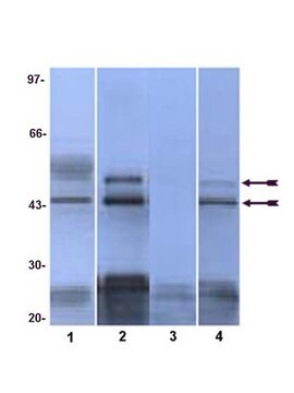
western blot: 1-2 μg/mL using using whole extracts of mouse NIH-3T3 cells

isotype

IgG2a

UniProt accession no.

shipped in

dry ice

storage temp.

−20°C

target post-translational modification

unmodified

Gene Information

human ... MAPK8(5599)
mouse ... Mapk8(26419)
rat ... Mapk8(116554)

General description

Anti-JNK antibody, Mouse monoclonal, (mouse IgG2a isotype) is derived from the hybridoma 1C2 produced by the fusion of mouse myeloma cells and splenocytes from BALB/c mice immunized with a recombinant protein. JNK1 (c-Jun N-terminal kinase), also known as MAPK8 (mitogen-activated protein kinase 8) is a member of the MAP kinase family. This gene is mapped to human chromosome 10q11.

Application

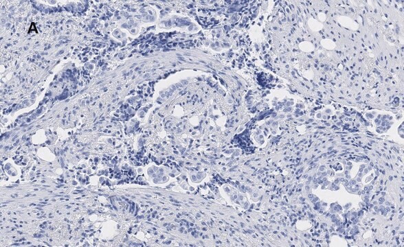
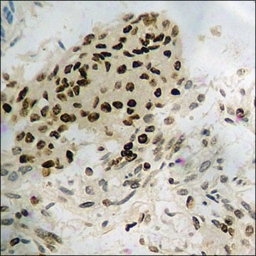
Anti-JNK antibody, Mouse monoclonal has been used in immunoblotting and enzyme linked immunosorbent assay (ELISA).

Biochem/physiol Actions

JNK1 (or MAPK8) is a serine threonine kinase that is a key regulator of cellular apoptosis. During the lack of NFkB stiμLation; persistent activation of JNK1 results in TNF-a induced cell death. However, JNK1 may also function as an apoptotic inhibitor. For instance; JNK1 interacts with WOX1 to inhibit apoptosis. Furthermore, JNK1 phosphorylates Bcl-2 to mediate starvation-induced autophagy, and also activates SIRT1 by phosphorylation during conditions of oxidative stress. JNK1 is activated by UV irradiation and oncoproteins and hence may be implicated in cancer propagation
Monoclonal Anti-JNK antibody is specific for human, mouse and rat JNK1 (43 kDa) and/or JNK2 (55 kDa).
JNK1 is also involved in UV (ultra violet) radiation induced apoptosis, which is thought to be related to cytochrome C-mediated cell death pathway.

Physical form

Solution in 0.01 M phosphate buffered saline, pH 7.4, containing 15 mM sodium azide.

Disclaimer

Unless otherwise stated in our catalog or other company documentation accompanying the product(s), our products are intended for research use only and are not to be used for any other purpose, which includes but is not limited to, unauthorized commercial uses, in vitro diagnostic uses, ex vivo or in vivo therapeutic uses or any type of consumption or application to humans or animals.

Not finding the right product?  

Try our Product Selector Tool.

Storage Class

10 - Combustible liquids

flash_point_f

Not applicable

flash_point_c

Not applicable


Certificates of Analysis (COA)

Search for Certificates of Analysis (COA) by entering the products Lot/Batch Number. Lot and Batch Numbers can be found on a product’s label following the words ‘Lot’ or ‘Batch’.

Already Own This Product?

Find documentation for the products that you have recently purchased in the Document Library.

Visit the Document Library

The Role of c-Jun N-Terminal Kinase (JNK) in Retinal Degeneration and Vision Loss
Kim BJ, et al.
Advances in Experimental Medicine and Biology, Adv(Exp), Med-Med (2018)
HSP70 Facilitates Memory Consolidation of Fear Conditioning through MAPK Pathway in the Hippocampus
Porto RR, et al.
Neuroscience, 375, 108-118 (2018)
JNK Signaling in Apoptosis
Dhanasekaran DN, et al.
Oncogene, 27(48), 6245-6245 (2008)
Molecular characterization of autophagic and apoptotic signaling induced by sorafenib in liver cancer cells
Rodriguez HMA, et al.
Journal of Cellular Physiology, 234(1), 692-708 (2019)
Esophageal squamous cell carcinomas with distinct invasive depth show different gene expression profiles associated with lymph node metastasis
Sato T, et al.
International Journal of Oncology, 28(5), 1043-1055 (2006)

Our team of scientists has experience in all areas of research including Life Science, Material Science, Chemical Synthesis, Chromatography, Analytical and many others.

Contact Technical Service