Skip to Content
MilliporeSigma
HomeSmall Molecules Analysis & QCOrganic impurity profiling of Valacyclovir using TLC

Organic impurity profiling method for Valacyclovir using TLC and following the United States Pharmacopoeia

Dr Sanjay Poman
Navi Mumbai, India

Abstract

This work introduces an analytical technique utilizing thin-layer chromatography (TLC) to analyze the organic contaminants in Valacyclovir Hydrochloride, an antiviral drug, following the guidelines set by the United States Pharmacopeia (USP). The method identifies related compounds D, E, F, and G using 0.25 mm Silica Gel 60 F254 TLC plates. The testing results exhibit excellent recovery and precision, with impurity levels falling below the acceptable requirements set by the United States Pharmacopeia (USP).

Section Overview

Introduction

Chemical structure of Valacyclovir Hydrochloride

Valacyclovir Hydrochloride

Valacyclovir is an antiviral drug. It shows the growth and spread of herpes virus to help the body fight the infection. Valacyclovir is used to treat infections caused by herpes viruses, including genital herpes, cold stores, and shingles (herpes zosters) in adults. It is also used to prevent cytomegalovirus following a kidney transplant in high-risk cases.1

The present study demonstrates the use of a thin layer chromatography (TLC) method (Table 1) for determination of valacyclovir organic impurities (related compounds D, E, G & F) according to the USP monograph method.2

Experimental

Table 1.Chromatographic conditions

Results

A thin-layer chromatography (TLC) plate, viewed under a wavelength of 254 nm after being sprayed with fluorescamine, displays a vibrant green color. On both the right and left edges of the plate, there are 9 red markings, starting from the bottom and labeled from 0.1 to 0.9, each placed at intervals of 0.1 cm. The plate reveals three distinct black bands, representing the impurities labeled D, E, and G. These bands stand out clearly against the green background, indicating the presence and separation of these impurities.

Figure 1a.Chromatograms at short (254 nm) wavelength.

A thin-layer chromatography (TLC) plate, viewed under a wavelength of 365 nm after being sprayed with fluorescamine, displays a blue color. On both the right and left edges of the plate, there are 9 yellow markings, starting from the bottom and labeled from 0.1 to 0.9, each placed at intervals of 0.1 cm. The plate reveals three distinct faded bands, representing the impurities labeled D, E, and G. These bands stand out clearly against the blue background, indicating the presence and separation of these impurities.

Figure 1b.Chromatograms at long (365 nm) wavelength.
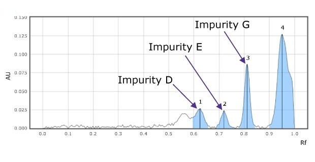
A TLC-densitogram obtained using a TLC analyzer for an impurity mix, scanned at 254 nm, shows the retention factor on the x-axis with major tick marks at intervals of 0.1 (from 0.1 to 0.9). The y-axis, depicting detector response in absorbance units (AU), has major tick marks starting at 0.025 and ending at 0.150, at intervals of 0.025. The densitogram displays four blue-colored peaks labeled 1, 2, 3, being impurities D,E, and G, and 4 being F from left to right, appearing between retention factor (Rf) values of 0.6 and 1.0.

Figure 2a.Densitograms of impurity mix at 254 nm for impurity D (1), E(2), and G(3).
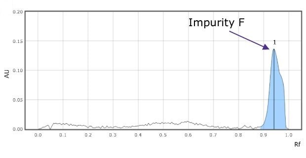
A TLC-densitogram obtained using a TLC analyzer for valacyclovir impurity F, scanned at 366 nm, shows the retention factor on the x-axis with major tick marks at intervals of 0.1 (from 0.1 to 0.9). The y-axis, depicting detector response in absorbance units (AU), has major tick marks starting at 0.05 and ending at 0.20, at intervals of 0.05. The densitogram displays one blue-colored peak labeled 1 being impurity F, appearing between retention factor (Rf) values of 0.9 and 1.0.

Figure 2b.Densitograms of impurity mix at 366 nm for impurity F (1) solution.
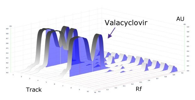
A 3-dimensional TLC-densitogram, obtained for the developed plate using a TLC analyzer and scanned at 254 nm, visualizes the separation and concentration of compounds. The x-axis on the flat rectangular surface shows the position of the spots, indicating where each compound starts on the plate. The y-axis displays the retention factor, showing how far each compound travels up the plate. The z-axis represents the detector's response in absorbance units (AU), with three peaks of varying heights rising from the surface. These peaks indicate the concentration of the compounds, with taller peaks signifying higher concentrations. The variation in peak heights across the distance traveled illustrates changes in concentration as the compounds move on the TLC plate

Figure 3.Densitogram of developed plate at 254 nm.

Table 2.Chromatographic data - standard solution. (*is quantitated by different procedure)
Table 3.Standard concentrations, relative concentration to sample solution and USP acceptance criteria for impurity content

Conclusion

The chromatogram for the impurity mixes shows three clearly separated spots (Figure 1 & 2).

The relative Rf values of the standard spots are close to the relative Rf value mentioned in the USP monograph (Table 2) and the valacyclovir sample shows less amount of impurities E, G, & F than the acceptance criteria values mentioned in the monograph (Table 3).

See more applications for Pharmaceutical Analysis and Quality Control

Related Products
Loading

References

1.
valACYclovir (Monograph). [Internet].[cited 08 Jul 2024]. Available from: https://www.drugs.com/monograph/valacyclovir
2.
Valacyclovir Hydrochloride. https://doi.org/10.31003/uspnf_m87560_04_01
Sign In To Continue

To continue reading please sign in or create an account.

Don't Have An Account?