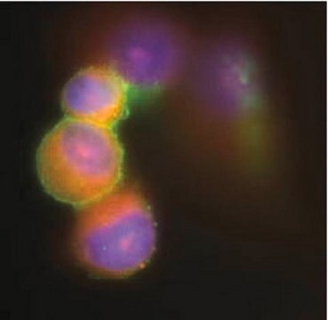
MABN754
Anti-PAH Antibody, clone 6H10.1
clone 6H10.1, from mouse
別名:
Phenylalanine-4-hydroxylase, PAH, Phe-4-monooxygenase
ログイン組織・契約価格を表示する
すべての画像(3)
About This Item
UNSPSCコード:
12352203
eCl@ss:
32160702
NACRES:
NA.41
クローン:
6H10.1, monoclonal
application:
IHC
WB
WB
化学種の反応性:
human
テクニック:
immunohistochemistry: suitable
western blot: suitable
western blot: suitable
citations:
おすすめの製品
詳細
PAH, also known as Phenylalanine-4-hydroxylase , Phe-4-monooxygenase, and encoded by the gene name PAH, belongs to the biopterin-dependent aromatic amino acid hydroxylase family. Phenylalanine hydroxylase is the rate-limiting enzyme of the metabolic pathway that degrades excess phenylalanine. Phenylalanine hydroxylase (PheOH, alternatively PheH or PAH) is an enzyme that catalyzes the hydroxylation of the aromatic side-chain of phenylalanine to generate tyrosine. PheOH is one of three members of the pterin-dependent amino acid hydroxylases, a class of monooxygenase that uses tetrahydrobiopterin (BH4, a pteridine cofactor) and a non-heme iron for catalysis. During the reaction, molecular oxygen is heterolytically cleaved with sequential incorporation of one oxygen atom into BH4 and phenylalanine substrate. PAH has been associated with Phenylketonuria PKU, an autosomal recessive inborn error of phenylalanine metabolism, due to severe phenylalanine hydroxylase deficiency. Additioanlly, PAH has been associated with Non-phenylketonuria hyperphenylalaninemia (Non-PKU HPA), a mild form of phenylalanine hydroxylase deficiency characterized by phenylalanine levels persistently below 600 mumol, which allows normal intellectual and behavioral development without treatment. Finally, PAH may play a role in the Hyperphenylalaninemia (HPA), a mildest form of phenylalanine hydroxylase deficiency. PAH is broadly expressed, with greatest levels in skeletal muscle followed by heart, brain, pancreas and testis.
免疫原
GST-tagged recombinant protein corresponding to human PAH.
アプリケーション
This Anti-PAH antibody is validated for use in WB, IH for the detection of PAH.
Western Blotting Analysis: 1.0 µg/mL from a representative lot detected PAH in 10 µg of human liver tissue lysate.
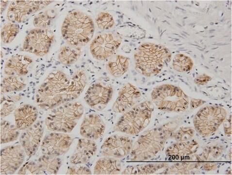
Immunohistochemistry Analysis: A 1:50-250 dilution from a representative lot detected PAH in human cerebral cortex and human liver tissue.
Immunohistochemistry Analysis: A 1:50-250 dilution from a representative lot detected PAH in human cerebral cortex and human liver tissue.
品質
Evaluated by Western Blotting in HepG2 cell lysate.
Western Blotting Analysis: 1.0 µg/mL of this antibody detected PAH in 10 µg of HepG2 cell lysate.
Western Blotting Analysis: 1.0 µg/mL of this antibody detected PAH in 10 µg of HepG2 cell lysate.
ターゲットの説明
~52 kDa observed
物理的形状
Format: Purified
その他情報
Concentration: Please refer to lot specific datasheet.
適切な製品が見つかりませんか。
製品選択ツール.をお試しください
保管分類コード
12 - Non Combustible Liquids
WGK
WGK 1
引火点(°F)
Not applicable
引火点(℃)
Not applicable
適用法令
試験研究用途を考慮した関連法令を主に挙げております。化学物質以外については、一部の情報のみ提供しています。 製品を安全かつ合法的に使用することは、使用者の義務です。最新情報により修正される場合があります。WEBの反映には時間を要することがあるため、適宜SDSをご参照ください。
Jan Code
MABN754:
試験成績書(COA)
製品のロット番号・バッチ番号を入力して、試験成績書(COA) を検索できます。ロット番号・バッチ番号は、製品ラベルに「Lot」または「Batch」に続いて記載されています。
ライフサイエンス、有機合成、材料科学、クロマトグラフィー、分析など、あらゆる分野の研究に経験のあるメンバーがおります。.
製品に関するお問い合わせはこちら(テクニカルサービス)

