おすすめの製品
由来生物
goat
品質水準
結合体
FITC conjugate
抗体製品の状態
affinity isolated antibody
抗体製品タイプ
secondary antibodies
クローン
polyclonal
形状
buffered aqueous solution
テクニック
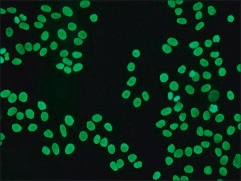
direct immunofluorescence: 1:32
保管温度
2-8°C
ターゲットの翻訳後修飾
unmodified
関連するカテゴリー
詳細
Human IgGs are glycoprotein antibodies that contain two equivalent light chains and a pair of identical heavy chains. IgGs have four distinct isoforms, ranging from IgG1 to IgG4. These antibodies regulate immunological responses to allergy and pathogenic infections. IgGs have also been implicated in complement fixation and autoimmune disorders
Anti-Human IgG (γ-chain specific), (F(ab′)2) fragment-FITC antibody is specific for human IgG when tested against purified human IgA, IgG, IgM, Bence Jones κ and λ myeloma proteins. The use of this product prevents background staining due to the presence of Fc receptors.
Anti-Human IgG (γ-chain specific), (F(ab′)2) fragment-FITC antibody is specific for human IgG when tested against purified human IgA, IgG, IgM, Bence Jones κ and λ myeloma proteins. The use of this product prevents background staining due to the presence of Fc receptors.
Immunoglobulin G (IgG) belongs to the immunoglobulin family and is a widely expressed serum antibody. The two heavy chains and two light chains of IgG are connected by a disulfide bond. It is a glycoprotein and mainly helps in immune defense. IgG is usually found as a monomer. IgG antibody subtype is the most abundant of serum immunoglobulins of the immune system. It is secreted by B cells and is found in blood and extracellular fluids. About 70 percent of the total immunoglobulin consists of IgG. Immunoglobulin G (IgG) participates in hypersensitivity type II and type III.
免疫原
Purified human IgG
アプリケーション
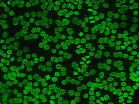
Anti-Human IgG (γ-chain specific), (F(ab′)2) fragment-FITC antibody is suitable for use in direct immunofluorescence (1:32).
Anti-Human IgG (γ-chain specific), F(ab′)2 fragment−FITC antibody has been used in immunofluorescence studies and flow cytometric crossmatch (FCXM).
物理的形状
0.01M PBS溶液(pH 7.4, 1%BSA, 15 mMアジ化ナトリウム含有)
免責事項
Unless otherwise stated in our catalog or other company documentation accompanying the product(s), our products are intended for research use only and are not to be used for any other purpose, which includes but is not limited to, unauthorized commercial uses, in vitro diagnostic uses, ex vivo or in vivo therapeutic uses or any type of consumption or application to humans or animals.
適切な製品が見つかりませんか。
製品選択ツール.をお試しください
保管分類コード
12 - Non Combustible Liquids
WGK
nwg
引火点(°F)
Not applicable
引火点(℃)
Not applicable
適用法令
試験研究用途を考慮した関連法令を主に挙げております。化学物質以外については、一部の情報のみ提供しています。 製品を安全かつ合法的に使用することは、使用者の義務です。最新情報により修正される場合があります。WEBの反映には時間を要することがあるため、適宜SDSをご参照ください。
Jan Code
F1641-2ML:
F1641-.5ML:
F1641-1ML:
F1641-VAR:
F1641PROC:
F1641-BULK:
試験成績書(COA)
製品のロット番号・バッチ番号を入力して、試験成績書(COA) を検索できます。ロット番号・バッチ番号は、製品ラベルに「Lot」または「Batch」に続いて記載されています。
この製品を見ている人はこちらもチェック
IgG
Encyclopedia of Immunology (1998)
Işın Sinem Bağcı et al.
Journal of biophotonics, 14(5), e202000509-e202000509 (2021-01-26)
Ex vivo confocal laser scanning microscopy (ex vivo CLSM) provides rapid, high-resolution imaging and immunofluorescence examinations of the excised tissues. We aimed to evaluate the applicability of ex vivo CLSM in histomorphological and direct immunofluorescence (DIF) examination of pemphigus vulgaris
I S Bağcı et al.
Journal of the European Academy of Dermatology and Venereology : JEADV, 33(11), 2123-2130 (2019-07-03)
Ex vivo confocal laser scanning microscopy (ex vivo CLSM) is a novel diagnostic method allowing rapid, high-resolution imaging of excised skin samples. Furthermore, fluorescent detection is possible using fluorescent-labelled antibodies. To assess the applicability of ex vivo CLSM in the
Local activation of the complement system in endoneurial microvessels of diabetic neuropathy
Rosoklija G B, et al.
Acta Neuropathologica, 99(1), 55-62 (2000)
Işın Sinem Bağcı et al.
Experimental dermatology, 30(5), 684-690 (2020-12-22)
Ex vivo confocal laser scanning microscopy (CLSM) offers real-time examination of excised tissue in reflectance, fluorescence and digital haematoxylin-eosin (H&E)-like staining modes enabling application of fluorescent-labelled antibodies. We aimed to assess the diagnostic performance of ex vivo CLSM in identifying
ライフサイエンス、有機合成、材料科学、クロマトグラフィー、分析など、あらゆる分野の研究に経験のあるメンバーがおります。.
製品に関するお問い合わせはこちら(テクニカルサービス)