추천 제품
생물학적 소스
rabbit
Quality Level
항체 형태
serum
항체 생산 유형
primary antibodies
클론
polyclonal
종 반응성
rat, human
제조업체/상표
Chemicon®
기술
ELISA: suitable
immunocytochemistry: suitable
immunohistochemistry (formalin-fixed, paraffin-embedded sections): suitable
radioimmunoassay: suitable
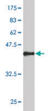
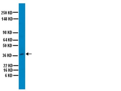
western blot: suitable
NCBI 수납 번호
UniProt 수납 번호
배송 상태
wet ice
타겟 번역 후 변형
unmodified
유전자 정보
human ... IBSP(3381)
rat ... Ibsp(24477)
일반 설명
Bone sialoprotein II is one of the major noncollagenous proteins in the extracellular matrix of bone. It is a phosphorylated glycoprotein with an approximate molecular weight of 70 kDa. The protein has been found in osteoblasts and osteocytes. Diseases concerning the bone turnover, pathological bone alterations as well as the rapid increase of osteoporosis make it necessary to establish new markers, Bone sialoprotein is discussed as a potential serum marker for monitoring bone remodelling.
특이성
Human and rat Bone sialoprotein (BSP). No cross reactivity with human osteopontin or osteonectin by RIA or ELISA.
Other species have not been tested
애플리케이션
Anti-Bone Sialoprotein II Antibody is an antibody against Bone Sialoprotein II for use in ELISA, IC, IH, IH(P), RIA & WB.
Western Blot
Immunohistochemistry (Paraffin) (see suggested protocol below)
Immunocytochemistry (1:100)
RIA
ELISA
Optimal working dilutions must be determined by the end user.
SUGGESTED STAINING PROCEDURE FOR RABBIT ANTI-BONE SIALOPROTEINAB1854
This protocol has been used successfully on 5 um, paraffin embedded sections from human fetal tibia, human fetal calvaria, human metastatic breast tumors to bone which were decalcified in 10% formic acid.
1. Dewaxing: Xylene 5 min. - Xylene 5 min. - Xylene 5 min.
2. Rehydration: 100% ethanol for 3 min. - 95% ethanol for 3 min. - 70% ethanol for 3 min.
3. Wash sections under running tap water for 5 minutes.
4. Wash sections in PBS.
5. Block endogenous peroxidase: 0.3% hydrogen peroxide in absolute methanol (2 mL of H2O2 30% in 198 mL methanol) for 30 minutes. (Cover the jar with foil, use stirring during incubation.)
6. Wash sections under running tap water for 10 minutes.
7. Mark tissue area with pap pen.
8. Cover the tissue area with 300 mL of blocking solution for 30 minutes at room temperature (blocking solution: 6 mL PBS, 0.3 gm BSA, 120 mL normal goat serum).
9. Shake off excess fluid (do not wash).
10. Incubate with primary antibody (AB1854 diluted 1:2,500 in PBS-T) at 2-8°C overnight in moist chamber.
11. Rinse with PBS-T, then wash 3 times in PBS-T each for 5 minutes at room temperature.
12. Incubate with biotinylated secondary antibody (for example Chemicon AP132B) for 30 minutes at room temperature.
13. Rinse with PBS-T, then wash in PBS-T 3 x 5 minutes at room temperature.
14. Incubate the sections with Vector ABC reagent for 30 minutes at room temperature. (ABC reagent: 2 drops of A and 2 drops of B, in 5 mL PBS, allow to stand 30 minutes at room temperature before use.)
15. Wash sections in PBS 3 x 5 minutes at room temperature.
16. Develop with DAB for 5-10 minutes.
17. Wash under running tap water.
18. Counter stain with Mayer′s HX for 45 seconds, wash in warm tap water.
19. Dehydrate: 70% ethanol - 95% ethanol - 100% ethanol, 1 min. each.
20. Wash in Xylene, mount, dry.
물리적 형태
Lyophilized. Resuspend in 100 μL aqua bidest. Contains no preservatives.
저장 및 안정성
Maintain at 2-8°C (lyophilized) or -20°C (reconstituted) for up to 6 months in undiluted aliquots. Avoid repeated freeze/thaw cycles.
법적 정보
CHEMICON is a registered trademark of Merck KGaA, Darmstadt, Germany
적합한 제품을 찾을 수 없으신가요?
당사의 제품 선택기 도구.을(를) 시도해 보세요.
Storage Class Code
10 - Combustible liquids
WGK
WGK 1
Flash Point (°F)
Not applicable
Flash Point (°C)
Not applicable
시험 성적서(COA)
제품의 로트/배치 번호를 입력하여 시험 성적서(COA)을 검색하십시오. 로트 및 배치 번호는 제품 라벨에 있는 ‘로트’ 또는 ‘배치’라는 용어 뒤에서 찾을 수 있습니다.
MG63 osteoblast-like cells exhibit different behavior when grown on electrospun collagen matrix versus electrospun gelatin matrix.
Tsai, SW; Liou, HM; Lin, CJ; Kuo, KL; Hung, YS; Weng, RC; Hsu, FY
Testing null
Y-C Huang et al.
Gene therapy, 12(5), 418-426 (2005-01-14)
Gene therapy approaches to bone tissue engineering have been widely explored. While localized delivery of plasmid DNA encoding for osteogenic factors is attractive for promoting bone regeneration, the low transfection efficiency inherent with plasmid delivery may limit this approach. We
Loss of MMP-2 in murine osteoblasts upregulates osteopontin and bone sialoprotein expression in a circuit regulating bone homeostasis.
Mosig, RA; Martignetti, JA
Disease models & mechanisms null
Cristina Correia et al.
PloS one, 6(12), e28352-e28352 (2011-12-14)
Tissue engineering provides unique opportunities for regenerating diseased or damaged tissues using cells obtained from tissue biopsies. Tissue engineered grafts can also be used as high fidelity models to probe cellular and molecular interactions underlying developmental processes. In this study
Majd Machour et al.
Advanced science (Weinheim, Baden-Wurttemberg, Germany), 9(34), e2200882-e2200882 (2022-10-20)
3D bioprinting holds great promise for tissue engineering, with extrusion bioprinting in suspended hydrogels becoming the leading bioprinting technique in recent years. In this method, living cells are incorporated within bioinks, extruded layer by layer into a granular support material
자사의 과학자팀은 생명 과학, 재료 과학, 화학 합성, 크로마토그래피, 분석 및 기타 많은 영역을 포함한 모든 과학 분야에 경험이 있습니다..
고객지원팀으로 연락바랍니다.


