
추천 제품


제품명
Anti-Tryptophan Hydroxylase/Tyrosine Hydroxylase/Phenylalanine Hydroxylase Antibody, clone PH8, clone PH8, Chemicon®, from mouse
생물학적 소스
mouse
Quality Level
항체 형태
purified immunoglobulin
항체 생산 유형
primary antibodies
클론
PH8, monoclonal
종 반응성
human
제조업체/상표
Chemicon®
기술
immunohistochemistry: suitable
immunoprecipitation (IP): suitable
western blot: suitable
동형
IgG1
NCBI 수납 번호
배송 상태
wet ice
타겟 번역 후 변형
unmodified
유전자 정보
human ... TPH1(7166)
일반 설명
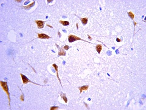
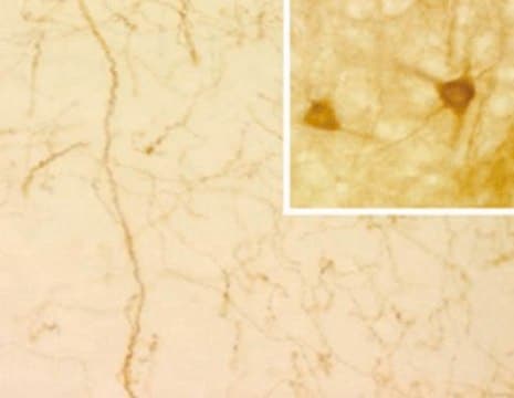
PH8 Antibody is a murine monoclonal antibody that binds a common epitope of Tryptophan hydroxylase (TRH), Tyrosine hydroxylase (TYH) and Phenylalanine hydroxylase (PAH).1 In the literature this monoclonal antibody is cited as PH8. Tyrosine hydroxylase (TYH) is the enzyme which converts tyrosine to dihydroxyphenylalanine (L-dopa), a precursor of the catecholamine neurotransmitters dopamine, noradrenaline and adrenaline. Tryptophan hydroxylase (TRH) is the enzyme that converts 5-hydroxytryptophan to serotonin. In fresh tissue the PH8 Antibody binds to TYH and TRH so is a marker for TYH-containing catecholaminergic neurons,2 and serotonergic neurons.3,4,5 Tryptophan hydroxylase (TRH) can be used as a marker for serotonin as it converts 5-hydroxy-tryptophan to serotonin. Serotonin is rapidly metabolised and is unable to be detected by anti-serotonin antibodies in post mortem tissue. In human tissue that has been formalin-fixed, due to a change in the antigenic determinant of tyrosine hydroxylase (TYH), PH8 Antibody will bind only tryptophan hydroxylase (TRH).3,4,5 The PH8 Antibody can therefore be used to specifically identify serotonergic neurons in fixed human tissue. Phenylalanine hydroxylase (PAH) can be detected in hepatic tissue sections using the PH8 Antibody.
애플리케이션
The PH8 Antibody can be used to identify dopaminergic and serotonergic neurons by immuno-histochemistry and for Western blot analysis, immunoprecipitation and immuno-histochemistry of TYH, PAH and TRH.
PROTOCOLS
Immunohistochemistry
The PH8 Antibody can be used for the immunohistochemical detection of dopaminergic and serotonergic neurons in human and rat brain stem tissue.
1. Tissue should be formalin fixed and stored in formalin prior to use for a minimum of five days.
2. Cryoprotect tissue using 30% sucrose in 0.1M Tris pH 7.4 buffer for 24-72 hours.
3. Cut using a sledge microtome to 50 μm thickness.
4. Wash the tissue samples using Tris buffer prior to commencing the staining procedure.
5. Treat the tissue samples for 3 x 15 minutes in 50% alcohol.
6. Treat the tissue samples for 20 minutes in 50% alcohol and 3% H2O2.
7. Treat tissue for 20 minutes with 10% normal horse serum in Tris buffer. This acts to block endogeneous H2O2 staining.
8. Dilute the PH8 Antibody in Tris buffer, add to the tissue samples and incubate for 1-3 days. Recommended dilutions are 1:2,000-1:10,000. At high antibody concentration all monoaminergic neurons are stained with no distinction between serotonegic and catecholominergic cells. By diluting the antibody concentrate, cell types are distinguishable due to variation in staining intensity. At lower concentrations (1:5,000-1:10,000 dilution) only serotonergic cells will stain.
9. Wash tissue (3 x 15 minutes), then add a biotinylated anti-mouse secondary antibody and incubate on an orbital shaker at room temperature (RT) for 1 hour.
10. Wash tissue (3 x 15 minutes) and incubate on an orbital shaker at RT for 1 hour with the tertiary complex (ELITE KIT, Vector, USA).
11. Wash tissue (3 x 15 minutes) and incubate on an orbital shaker at RT for 10 minutes with Tris buffered diamino-benzidine substrate. Add 0.1% H2O2 and incubate on an orbital shaker at RT for a further 5 minutes.
12. Mount tissue onto gelatinised slides and allow to dry prior to microscopy.
This Anti-Tryptophan Hydroxylase/Tyrosine Hydroxylase/Phenylalanine Hydroxylase Antibody, clone PH8 is validated for use in IHC, IP, WB for the detection of Tryptophan Hydroxylase/Tyrosine Hydroxylase/Phenylalanine Hydroxylase.
물리적 형태
Format: Purified
The immunoglobulin fraction has been purified by Protein G chromatography. 500?g liquid protein at 2mg/mL in phosphate buffered saline (PBS). The product is supplied sterile-filtered through a 0.22 micron filter
저장 및 안정성
The PH8 Antibody is shipped liquid at ambient temperature. Liquid material is stable for at up to 6 months when stored at 2-8°C.
법적 정보
CHEMICON is a registered trademark of Merck KGaA, Darmstadt, Germany
적합한 제품을 찾을 수 없으신가요?
당사의 제품 선택기 도구.을(를) 시도해 보세요.
Storage Class Code
12 - Non Combustible Liquids
WGK
WGK 2
Flash Point (°F)
Not applicable
Flash Point (°C)
Not applicable
시험 성적서(COA)
제품의 로트/배치 번호를 입력하여 시험 성적서(COA)을 검색하십시오. 로트 및 배치 번호는 제품 라벨에 있는 ‘로트’ 또는 ‘배치’라는 용어 뒤에서 찾을 수 있습니다.
Fiona M Bright et al.
Journal of neuropathology and experimental neurology, 76(10), 864-873 (2017-09-20)
Serotonin (5-hydroxytryptamine [5-HT]) neurons in the medulla oblongata project extensively to key autonomic and respiratory nuclei in the brainstem and spinal cord regulating critical homeostatic functions. Multiple abnormalities in markers of 5-HT function in the medulla in sudden infant death
Brainstem deficiency of the 14-3-3 regulator of serotonin synthesis: a proteomics analysis in the sudden infant death syndrome.
Broadbelt, KG; Rivera, KD; Paterson, DS; Duncan, JR; Trachtenberg, FL; Paulo, JA; Stapels et al.
Molecular and Cellular Proteomics null
Yuefeng Lu et al.
Anatomical record (Hoboken, N.J. : 2007), 293(11), 1954-1965 (2010-08-25)
Several lines of evidence have implicated a direct reciprocal interaction between serotonin and nitric oxide (NO). The goal of this investigation was, therefore, to examine the coexpression of tryptophan hydroxylase (TPH; the rate limiting enzyme for the synthesis of serotonin)
Rana A Eser et al.
Journal of neuropathology and experimental neurology, 77(2), 149-161 (2018-01-06)
The brainstem nuclei of the reticular formation (RF) are critical for regulating homeostasis, behavior, and cognition. RF degenerates in tauopathies including Alzheimer disease (AD), progressive supranuclear palsy (PSP), and corticobasal degeneration (CBD). Although the burden of phopho-tau inclusion is high
Kathleen A Sluka et al.
Pain reports, 5(6), e857-e857 (2020-12-10)
Regular physical activity/exercise is an effective nonpharmacological treatment for individuals with chronic pain. Central inhibitory mechanisms, involving serotonin and opioids, are critical to analgesia produced by regular physical activity. The rostral ventromedial medulla (RVM) sends projections to the spinal cord
자사의 과학자팀은 생명 과학, 재료 과학, 화학 합성, 크로마토그래피, 분석 및 기타 많은 영역을 포함한 모든 과학 분야에 경험이 있습니다..
고객지원팀으로 연락바랍니다.