94433
β-Galactosidase stain
동의어(들):
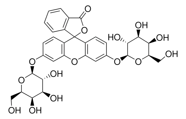
6′-(Diethylamino)-4′-(fluoromethyl)spiro[isobenzofuran-1(3H),9′-[9H]xanthen]-3′-yl β-D-galactopyranoside, SPiDER-βGal, SPiDER-beta Gal, (2S,3R,4S,5R,6R)-2-{[3’-(Diethylamino)-5’-(fluoromethyl)-3H-spiro(isobenzofuran-1,9’-xanthen)-6’-yl]oxy}-6-(hydroxymethyl)tetrahydro-2H-pyran-3,4,5-triol
About This Item
추천 제품
양식
solid
Quality Level
저장 온도
2-8°C
SMILES string
O[C@@H]1[C@@H](O)[C@H](OC(C=C2)=C(CF)C3=C2C4(C(C=CC=C5)=C5CO4)C(C=CC(N(CC)CC)=C6)=C6O3)O[C@H](CO)[C@@H]1O
InChI
1S/C31H34FNO8/c1-3-33(4-2)18-9-10-21-24(13-18)39-29-19(14-32)23(40-30-28(37)27(36)26(35)25(15-34)41-30)12-11-22(29)31(21)20-8-6-5-7-17(20)16-38-31/h5-13,25-28,30,34-37H,3-4,14-16H2,1-2H3/t25-,26+,27+,28-,30-,31?/m1/s1
InChI key
SVEDIEUQTRCORD-JGANWENXSA-N
일반 설명
To overcome these issues, Urano, Kamiya and co-workers have successfully developed a stain ideally possesses cell-permeability and the ability to retain in intracellular region.1)
By the enzymatic reaction, the β-Galactosidase stain immediately forms a quinone methide that acts as electrophile when proteins containing nucleophilic functional groups nearby the molecules. By the probe undergoes the reaction with a protein, the conjugates become fluorescent compounds. Thus, it allows a single-cell analysis because it does self-immobilizing to the intracellular proteins.
Exciation Maximum: 530 nm (± 10 nm)
Emission Maximum: 550 nm (± 10 nm)
Reference:
1) Y. Urano, M. Kamiya, T. Doura, WO 2015174460, A1, (19, November, 2015).
Find more infomation here 94433
애플리케이션
Add 35 μl of DMSO to a tube of stain solution (20 μg) and dissolve it with pipetting.
*Store the stock solution at -20°C.
Preparation of 1 μmol/l stain working solution
Dilute the stain DMSO stock solution with Hanks′ HEPES buffer to prepare 1 μmol/l stain working solution.
*Hanks′ HEPES buffer is recommended to maintain cell condition.
General protocol:
staining
1. Prepare cells for the assay.
2. Discard the culture medium and wash the cells with Hanks′ HEPES buffer twice.
3. Add an appropriate volume of stain working solution.
4. Incubate at 37oC for 15 minutes.
5. Observe the cells under a fluorescence microscope or by a flow cytometer.
*After staining, the cells can be observed even without washing. However, you can wash it as needed.
Storage Class Code
11 - Combustible Solids
WGK
WGK 3
자사의 과학자팀은 생명 과학, 재료 과학, 화학 합성, 크로마토그래피, 분석 및 기타 많은 영역을 포함한 모든 과학 분야에 경험이 있습니다..
고객지원팀으로 연락바랍니다.


