Skip to Content
MilliporeSigma
All Photos(5)

Key Documents

05-373

Sigma-Aldrich

Anti-Cyclin B1 Antibody, clone GNS3 (8A5D12)

clone GNS3 (8A5D12), Upstate®, from mouse

Synonym(s):

G2/mitotic-specific cyclin B1, cyclin B1

Sign Into View Organizational & Contract Pricing


About This Item

UNSPSC Code:
12352203
eCl@ss:
32160702
NACRES:
NA.41

biological source

mouse

Quality Level

antibody form

purified antibody

antibody product type

primary antibodies

clone

GNS3 (8A5D12), monoclonal

species reactivity

human, mouse

packaging

antibody small pack of 25 μg

manufacturer/tradename

Upstate®

technique(s)

immunohistochemistry: suitable
immunoprecipitation (IP): suitable
western blot: suitable

isotype

IgG

NCBI accession no.

UniProt accession no.

shipped in

dry ice

target post-translational modification

unmodified

Gene Information

human ... CCNB1(891)
mouse ... Ccnb1(268697)

General description

G2/mitotic-specific cyclin-B1 (UniProt: P14635; also known as Cyclin B1) is encoded by the CCNB1 (also known as CCNB) gene (Gene ID: 891) in human. Cyclins are the regulatory subunits of the cell cycle-dependent kinases (CDKs) that are responsible for the phosphorylation of several cellular targets. Cyclins contain the nuclear localization sequence (NLS) that help move CDKs into the nucleus. They also contain PEST (Pro, Glu, Ser, and Thr) sequences that target them for degradation by the ubiquitin-proteasomal pathway. Once the CDKs have completed their role, they undergo a rapid programmed proteolysis via ubiquitin-mediated delivery to the proteasome complex. Cyclin B1, a regulatory protein involved in mitosis, complexes with CDK1 to form the maturation-promoting factor (MPF). It is shown to be essential for the control of the cell cycle at the G2/M (mitosis) transition. It accumulates steadily during G2 phase and is abruptly destroyed at mitosis. Hence, the cyclin B1-CDK1 complex is considered to be a key regulator for mitotic entry. This complex phosphorylates a number of proteins prior to mitotic entry. Although five serine phosphorylation sites are described for cyclin B1, (Ser 116, 126, 128, 133, and 147), serine 133 phosphorylation by PLK1 regulates the entry of Cyclin B1-CDK1 complex into the nucleus during prophase. At the end of mitosis, cyclin B1 is rapidly removed by a ubiquitin ligase (anaphase-promoting complex/cyclosome) loaded with the targeting subunit CDC20. Activated cyclin B1-CDK1 complex is reported to catalyze its own destruction by stimulating the activity of APC. (Ref.: Van Zon, W., et al. (2010). J. Cell. Biol. 190(4); 587-602; Yuan, J., et al. (2004). Oncogene 23(34); 5843-5852).

Specificity

In addition to human, weak species cross-reactivity was observed with mouse.
This antibody is specific for human cyclin B1, Mr 58 kDa. Does not cross-react with cyclin A or cyclin D.

Application

Immunoprecipitation:
2 μg of a previous lot immunoprecipitated human cyclin B1 and cdc2 kinase from 500 μg of A431 RIPA lysate as determined by a subsequent immunoblot of the immunoprecipitate using anti-cdk1/cdc2 (PSTAIR), (Catalog # 06-923).
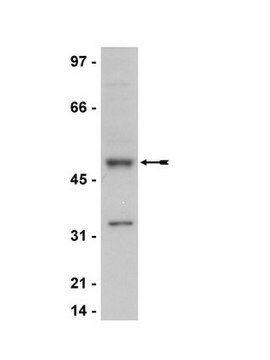
Western Blotting Analysis: 0.1 mg/mL of this antibody detected Cyclin B1 in A431 cell lysate.
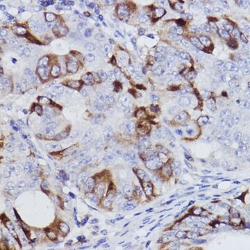
Immunohistochemistry (Paraffin) Analysis: A 1:250 and 1:50 dilutions of this antibody detected Cyclin B1 in Human tonsil and Human bone marrow tissue sections, respectively.

Quality

Routinely evaluated by western blot analysis on RIPA lysate from human A431 carcinoma cells.

Western Blot Analysis:
0.5-2 μg/mL of this lot detected cyclin B1 in RIPA lysates from human A431 carcinoma cells.

Target description

58 kDa

Linkage

Replaces: 04-220

Physical form

Format: Purified
Protein G purified mouse IgGs in storage buffer containing 0.1 M Tris-glycine, pH 7.4, 0.15 M NaCl, 0.05% sodium azide. Frozen at -20°C.

Analysis Note

Control
Positive Antigen Control: Catalog #12-301, non-stimulated A431 cell lysate. Add 2.5µL of 2-mercaptoethanol/100µL of lysate and boil for 5 minutes to reduce the preparation. Load 20µg of reduced lysate per lane for minigels.

Other Notes

Concentration: Please refer to the Certificate of Analysis for the lot-specific concentration.

Legal Information

UPSTATE is a registered trademark of Merck KGaA, Darmstadt, Germany

Not finding the right product?  

Try our Product Selector Tool.


Certificates of Analysis (COA)

Search for Certificates of Analysis (COA) by entering the products Lot/Batch Number. Lot and Batch Numbers can be found on a product’s label following the words ‘Lot’ or ‘Batch’.

Already Own This Product?

Find documentation for the products that you have recently purchased in the Document Library.

Visit the Document Library

Antiproliferation and radiosensitization of caffeic acid phenethyl ester on human medulloblastoma cells.
Lin, Yi-Hsien, et al.
Cancer Chemotherapy and Pharmacology, 57, 525-532 (2006)
Valosin-containing protein is a multi-ubiquitin chain-targeting factor required in ubiquitin-proteasome degradation.
R M Dai, C C Li
Nature Cell Biology null
Matthew S Fuller et al.
Journal of virology, 91(14) (2017-04-28)
Replication of minute virus of mice (MVM) induces a sustained cellular DNA damage response (DDR) which the virus then exploits to prepare the nuclear environment for effective parvovirus takeover. An essential aspect of the MVM-induced DDR is the establishment of
Kalyan Dulla et al.
Molecular & cellular proteomics : MCP, 9(6), 1167-1181 (2010-01-26)
Reversible protein phosphorylation is a key regulatory mechanism of mitotic progression. Importantly, protein kinases themselves are also regulated by phosphorylation-dephosphorylation processes; hence, phosphorylation dynamics of kinases hold a wealth of information about phosphorylation networks. Here, we investigated the site-specific phosphorylation
Phosphorylation-dependent binding of cyclin B1 to a Cdc6-like domain of human separase.
Boos, D; Kuffer, C; Lenobel, R; Korner, R; Stemmann, O
The Journal of Biological Chemistry null

Our team of scientists has experience in all areas of research including Life Science, Material Science, Chemical Synthesis, Chromatography, Analytical and many others.

Contact Technical Service