05-665
Anti-Active-β-Catenin (Anti-ABC) Antibody, clone 8E7
clone 8E7, Upstate®, from mouse
Synonym(s):
Anti-Anti-CTNNB, Anti-Anti-EVR7, Anti-Anti-MRD19, Anti-Anti-NEDSDV, Anti-Anti-armadillo
About This Item
Recommended Products
biological source
mouse
Quality Level
antibody form
purified immunoglobulin
antibody product type
primary antibodies
clone
8E7, monoclonal
species reactivity
human, rat, mouse
packaging
antibody small pack of 25 μg
manufacturer/tradename
Upstate®
technique(s)
flow cytometry: suitable
immunocytochemistry: suitable
immunohistochemistry (formalin-fixed, paraffin-embedded sections): suitable
western blot: suitable
isotype
IgG1κ
NCBI accession no.
UniProt accession no.
shipped in
ambient
target post-translational modification
unmodified
Gene Information
human ... CTNNB1(1499)
mouse ... Ctnnb1(12387)
rat ... Ctnnb1(84353)
Related Categories
General description
When β-catenin was sequenced it was found to be a member of the armadillo family of proteins. These proteins have multiple copies of the so-called armadillo repeat domain which is specialized for protein-protein binding. An increase in β-catenin production has been noted in those people who have Basal Cell Carcinoma and leads to the increase in proliferation of related tumors. When β-catenin is not associated with cadherins and α-catenin, it can interact with other proteins such as Catenin Beta Interacting Protein 1 (ICAT) and adenomatosis polyposis coli (APC).
Recent evidence suggests that β-catenin plays an important role in various aspects of liver biology including liver development (both embryonic and postnatal), liver regeneration following partial hepatectomy. HGF-induced hepatpomegaly, liver zonation, and pathogenesis of liver cancer.
Specificity
Immunogen
Application
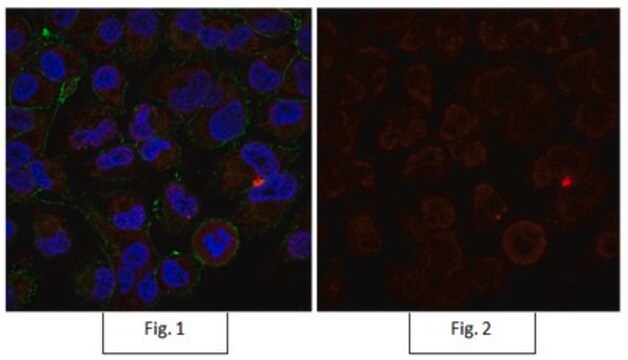
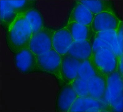
This antibody has also been reported by an independent laboratory to show positive immunostaining for beta-catenin in LiCl-treated 293T cells fixed with methanol (Staal, Frank J. T., 2002).
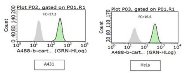
Flow Cytometry: This antibody was used in flow cytometry at an optimal 1 μg/mL concentration.
Immunohistochemistry: This antibody was used in immunohistochemistry on a colorectal carcinoma tissue array at a 1:300 dilution.
This antibody has also been reported by an independent laboratory to detect beta-catenin in mouse embryo sections (Van Noort, M., 2002).
Epigenetics & Nuclear Function
Transcription Factors
Quality
Western Blot Analysis: 0.2-2 µg/mL of this antibody detected β-catenin in RIPA lysates from A431 cells.
Target description
Physical form
Storage and Stability
Handling Recommendations: Upon receipt, and prior to removing the cap, centrifuge the vial and gently mix the solution.
Analysis Note
Positive Antigen Control: Catalog #12-301, non-stimulated A431 cell lysate. Add 2.5µL of 2-mercaptoethanol/100µL of lysate and boil for 5 minutes to reduce the preparation. Load 20µg of reduced lysate per lane for minigels.
Legal Information
Disclaimer
Not finding the right product?
Try our Product Selector Tool.
recommended
Storage Class
12 - Non Combustible Liquids
wgk_germany
WGK 1
flash_point_f
Not applicable
flash_point_c
Not applicable
Certificates of Analysis (COA)
Search for Certificates of Analysis (COA) by entering the products Lot/Batch Number. Lot and Batch Numbers can be found on a product’s label following the words ‘Lot’ or ‘Batch’.
Already Own This Product?
Find documentation for the products that you have recently purchased in the Document Library.
Customers Also Viewed
Our team of scientists has experience in all areas of research including Life Science, Material Science, Chemical Synthesis, Chromatography, Analytical and many others.
Contact Technical Service


