Skip to Content
MilliporeSigma
All Photos(2)

Key Documents

07-1210

Sigma-Aldrich

Anti-IP3 Receptor Antibody

from rabbit

Synonym(s):

Inositol 1,4,5-Trisphosphate Receptor

Sign Into View Organizational & Contract Pricing


About This Item

UNSPSC Code:
12352203
eCl@ss:
32160702
NACRES:
NA.41

biological source

rabbit

Quality Level

antibody form

purified immunoglobulin

antibody product type

primary antibodies

clone

polyclonal

species reactivity

pig, human, bovine, rabbit, mouse, canine, dog, rat

technique(s)

ELISA: suitable
immunofluorescence: suitable
immunohistochemistry: suitable (paraffin)
immunoprecipitation (IP): suitable
radioimmunoassay: suitable
western blot: suitable

isotype

IgG

NCBI accession no.

UniProt accession no.

shipped in

dry ice

target post-translational modification

unmodified

Gene Information

human ... ITPR1(3708)

General description

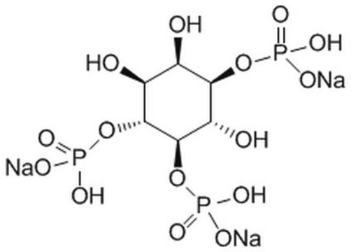
The InsP3 (inositol 1,4,5-trisphosphate) Receptor family are intracellular Ca2+ channels activated by the second messenger InsP3. There are at least three types of receptor, with distinct tissue-specific expression profiles and subcellular localizations; it is thought that the exact subtype profile conveys precise Ca2+ release responses. Release of Ca2+ from intracellular stores is the trigger for many cellular responses, including muscle contraction. Receptor activity is subject to feedback regulation by Ca2+ concentration, as high cytosolic Ca2+ concentrations shut down the channel. Ca2+ release is augmented by phosphorylation of the Receptor by PKA and PKG.

Specificity

Reactivity with other species has not been determined.
Reacts with the C-terminal cytoplasmic domain of IP3 receptors Type 1, 2, and 3. IP3 receptors have been shown to play an important role in the regulation of Ca2+ release from internal storage sites during cell activation. AB1622 reacts with IP3 receptor (MW approx. 260 kDa) isolated from a variety of cell types (e.g. lymphocytes, macrophages, granulocytes, fibroblasts, epithelial, endothelial cells, skeletal muscle, cardiac muscle and brain tissues).

Immunogen

Epitope: C-terminus
Synthetic peptide (KDSTEYTGPESYV) corresponding to the C-terminus of human IP3 Receptor type 1.

Application

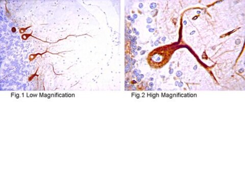
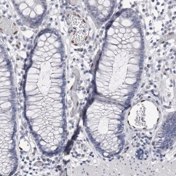
Immunohistochemisty (paraffin): IP3 Receptor staining in rat cerebellum, tissue was pretreated with citrate buffer, pH 6.0. A previous lot of this antibody was diluted to 1:100, using IHC-Select detection with HRP-DAB.

Immunofluorescence: 1:20 - 1:200 dilution from a previous lot. Light fixation, 2% PFA, 90% ethanol fixation; Visualization by confocal microscopy is required, as detection by standard fluorescent microscopy will not be adequate to detect the IP3R. Due to the intensity of confocal lasers, use of an anti-fading agent, such as DABCO, is strongly recommended.

ELISA: 1:2,000 dilution of a previous was used in ELISA.

Immunoprecipitation: 1:200 dilution of a previous lot was used in Immunoprecipitation.

Flow cytometry: 1:1,000 dilution of a previous lot was used in Flow Cytometry.

Radioimmunoassay: 1:10,000 dilution of a previous lot was used in Radioimmunoassay.

Optimal working dilutions must be determined by the end user.
Research Category
Signaling
Research Sub Category
GPCR, cAMP/cGMP & Calcium Signaling
This Anti-IP3 Receptor Antibody is validated for use in WB, IF, IH(P), ELISA, IP, RIA for the detection of IP3 Receptor.

Quality

Routinely evaluated by Western Blot on RAW 264 lysates.

Western Blot Analysis:
1:500-1,000 dilution of this lot detected IP3 Receptor on 10 μg of RAW 264 lysates.

Target description

~260 kDa

Linkage

Replaces: AB1622

Physical form

Format: Purified
Purified
Purified rabbit polyclonal IgG in buffer containing 10 mM sodium phosphate, 150 mM NaCl, and 0.01% sodium azide, pH 7.5.

Storage and Stability

Stable for 1 year at -20°C in undiluted aliquots from date of receipt.
Handling Recommendations: Upon first thaw, and prior to removing the cap, centrifuge the vial and gently mix the solution. Aliquot into microcentrifuge tubes and store at -20°C. Avoid repeated freeze/thaw cycles, which may damage IgG and affect product performance.

Analysis Note

Control
RAW264 cell lysate

Other Notes

Concentration: Please refer to the Certificate of Analysis for the lot-specific concentration.

Disclaimer

Unless otherwise stated in our catalog or other company documentation accompanying the product(s), our products are intended for research use only and are not to be used for any other purpose, which includes but is not limited to, unauthorized commercial uses, in vitro diagnostic uses, ex vivo or in vivo therapeutic uses or any type of consumption or application to humans or animals.

Not finding the right product?  

Try our Product Selector Tool.

Storage Class

12 - Non Combustible Liquids

wgk_germany

WGK 2

flash_point_f

Not applicable

flash_point_c

Not applicable


Certificates of Analysis (COA)

Search for Certificates of Analysis (COA) by entering the products Lot/Batch Number. Lot and Batch Numbers can be found on a product’s label following the words ‘Lot’ or ‘Batch’.

Already Own This Product?

Find documentation for the products that you have recently purchased in the Document Library.

Visit the Document Library

Hongwei Dou et al.
The journal of histochemistry and cytochemistry : official journal of the Histochemistry Society, 52(10), 1377-1384 (2004-09-24)
The endolymph in the endolymphatic sac (ES) is acidic (pH 6.6-7). Maintaining this acidic lumen is believed to be important for the normal function of the ES. The acid-base regulation mechanisms of the ES are unknown. Here we investigated the
The involvement of the cytoskeleton in regulating IP3 receptor-mediated internal Ca2+ release in human blood platelets.
Bourguignon, L Y, et al.
Cell Biology International, 17, 751-758 (1993)
L Y Bourguignon et al.
The Journal of biological chemistry, 268(10), 7290-7297 (1993-04-05)
Mouse T-lymphoma cells contain a unique type of internal vesicle which bands at the relatively light density of 1.07 g/cc. These vesicles do not contain any detectable Golgi, endoplasmic reticulum, plasma membrane, or lysosomal marker protein activities. Binding of [3H]inositol
Identification of an IP3 receptor in endothelial cells.
Bourguignon, L Y, et al.
Journal of Cellular Physiology, 159, 29-34 (1994)
Egor A Turovsky et al.
International journal of molecular sciences, 22(10) (2021-06-03)
Various types of cells demonstrate ubiquitous rhythmicity registered as simple and complex Ca2+-oscillations, spikes, waves, and triggering phenomena mediated by G-protein and tyrosine kinase coupled receptors. Phospholipase C/IP3-receptors (PLC/IP3R) and endothelial NO-synthase/Ryanodine receptors (NOS/RyR)-dependent Ca2+ signaling systems, organized as multivariate

Our team of scientists has experience in all areas of research including Life Science, Material Science, Chemical Synthesis, Chromatography, Analytical and many others.

Contact Technical Service