Skip to Content
MilliporeSigma
All Photos(3)

Key Documents

17-10060

Sigma-Aldrich

ChIPAb+ NFκB p65 (RelA) - ChIP Validated Antibody and Primer Set

from mouse

Synonym(s):

Chip Antibody and primer set, Nuclear factor NF-kappa-B p65 subunit ChIP, Transcription factor p65 ChIP, Transcription factor p65, Nuclear factor NF-kappa-B p65 subunit, Nuclear factor of kappa light polypeptide gene enhancer in B-cells 3

Sign Into View Organizational & Contract Pricing


About This Item

UNSPSC Code:
12352203
eCl@ss:
32160702
NACRES:
NA.41
clone:
monoclonal
application:
ChIP
IP
WB
species reactivity:
rat, human
technique(s):
ChIP: suitable
immunoprecipitation (IP): suitable
western blot: suitable
citations:
20

biological source

mouse

Quality Level

clone

monoclonal

species reactivity

rat, human

species reactivity (predicted by homology)

mouse

manufacturer/tradename

ChIPAb+
Upstate®

technique(s)

ChIP: suitable
immunoprecipitation (IP): suitable
western blot: suitable

isotype

IgG3

NCBI accession no.

UniProt accession no.

shipped in

dry ice

General description

All ChIPAb+ antibodies are individually validated for chromatin precipitation, every lot, every time. Each ChIPAb+ antibody set includes control primers (tested every lot by qPCR) to biologically validate your IP results in a locus-specific context. The qPCR protocol and primer sequences are provided, allowing researchers to validate ChIP protocols when using our antibody in their chromatin context. Each set also includes a negative control antibody to ensure specificity of the ChIP reaction.
The ChIPAb+ NFκB p65 (RelA) set includes the NFκB p65 (RelA) antibody, a negative control normal mouse IgG, and qPCR primers which amplify a 299 bp region of human IκBα promoter. The NFκB p65 (RelA) and negative controls are supplied in a scalable "per ChIP" reaction size and can be used to functionally validate the precipitation of NFκB p65 (RelA)-associated chromatin.
The transcription factor NFkappaB (Nuclear Factor kappa B) is involved in the expression and regulation of a number of important cellular and physiological processes such as growth, development, apoptosis, immune and inflammatory response, and activation of various viral promoters including human immunodeficiency virus long terminal repeats. NFkappaB represents a group of structurally related and evolutionarily conserved proteins related to the proto-oncogene c-Rel with five members in mammals that include Rel (cRel), RelA (p65), RelB, NFkappaB1 (p50 and its precursor p105), and NFkappaB2 (p52 and its precursor p100). NFkappaB/Rel proteins exist as homo- or heterodimers to form transcriptionally competent or repressive complexes. Although most NFkappaB dimers are activators of transcription, the p50/50 and p52/52 homodimers can repress the transcription of their target genes. The p50/p65 heterodimer of NFkappaB is the most abundant in cells.

Specificity

This antibody recognizes an epitope overlapping the nuclear location signal (NLS) of the p65 subunit of the NFkB heterodimer. Thus it selectively binds to the activated form of NFkB.

Immunogen

Epitope: NLS of p65 subunit
Peptide corresponding to human p65 coupled to BSA.

Application

Chromatin Immunoprecipitation:
Representative lot data.
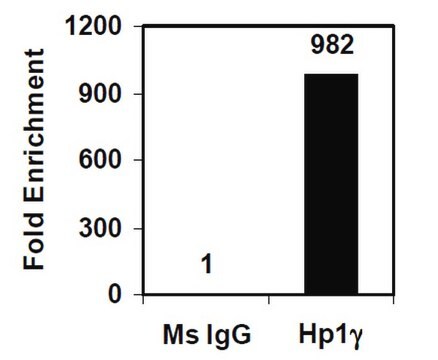
Sonicated chromatin prepared from serum starved, TNFα-treated (20 ng/mL, 30 min) 293 cells (~3 X 10E6 cell equivalents per IP) were subjected to chromatin immunoprecipitation using 4 µg of Normal Mouse IgG or 4 µg of Anti-NFκB p65 (RelA) and the Magna ChIP A Kit (Cat. # 17-610).
Successful immunoprecipitation of NFκB p65 (RelA) associated DNA fragments was verified by qPCR using ChIP Primers, IĸBα promoter as a positive locus, and β-Actin promoter primers as a negative locus (Please see figures). Data is presented as percent input of each IP sample relative to input chromatin for each amplicon and ChIP sample as indicated.
Please refer to the EZ-Magna ChIP A (Cat. # 17-409) or EZ-ChIP (Cat. # 17-371) protocol for experimental details.


Western Blot Analysis:
Representative lot data.
Huvec lysate (Lane 1), L6 lysate (Lane 2) and PC12 lysate (Lane 3) were resolved by electrophoresis, transferred to PVDF membrane and probed with anti-NFκB p65 (RelA) (1:500 dilution).
Proteins were visualized using a goat anti-mouse secondary antibody conjugated to HRP and a chemiluminescence detection
system.
Arrows indicates protein NFκB p65 (RelA) (~65 kDa) (Please see figures).
A non-specific band may be seen at ~230 kDa in L6 lysate.

Immunofluorescence: A 1-10 μg/mL concentration of a previous lot was used in immunofluorescence.

Immunohistochemistry (paraffin sections): A 5-10 μg/mL (APAAP)
concentration of a previous lot was used in immunohistochemistry.

Immunohistochemistry (frozen sections): A 5-10 μg/mL (APAAP)
concentration of a previous lot was used in immunohistochemistry.

Electrophoretic Mobility Shift Assay (EMSA): A 0.5-1 μg/mL concentration of a previous lot was used in shift assay.

Flow Cytometry: A previous lot of this antibody was used in flow cytometry.

Optimal working dilutions must be determined by end user.
Research Category
Epigenetics & Nuclear Function
Research Sub Category
Transcription Factors
This ChIPAb+ NFκB p65 (RelA) -ChIP Validated Antibody & Primer Set conveniently includes the antibody & the specific control PCR primers.

Packaging

25 assays per set. Recommended use: ~4 μg of antibody per chromatin immunoprecipitation (dependent upon biological context).

Quality

Chromatin Immunoprecipitation:
Sonicated chromatin prepared from serum starved, TNFα-treated (20 ng/mL, 30 min) 293 cells (~3 X 10E6 cell equivalents per IP) were subjected to chromatin immunoprecipitation using 4 µg of either Normal Mouse IgG or 4 µg of Anti-NFκB p65 (RelA) and the Magna ChIP® A Kit (Cat. # 17-610).
Successful immunoprecipitation of NFκB p65 (RelA) associated DNA fragments was verified by qPCR using ChIP Primers, IĸBα promoter (Please see figures).
Please refer to the EZ-Magna ChIP A (Cat. # 17-408) or EZ-ChIP (Cat. # 17-371) protocol for experimental details.

Target description

~65 kDa

Physical form

Anti-NFκB p65 (RelA) (mouse monoclonal). One vial containing 100 µg of protein A purified monoclonal IgG3 in a total volume of 143 uL in a buffer containing 0.02 M phosphate buffer, 0.25 M NaCl, pH 7.6 with 0.09% sodium azide, before the addition of glycerol to 30%. Store at -20°C.

Nornal Mouse IgG. One vial containing 125 µg of purified mouse IgG in 125 µL of storage buffer containing 0.1% sodium azide. Store at -20°C.

ChIP Primers, IĸBα promoter. One vial containing 75 μL of 5 μM of each primer specific for human IĸBα promoter. Store at -20°C.
FOR: GAC GAC CCC AAT TCA AAT CG
REV: TCA GGC TCG GGG AAT TTC C
Format: Purified
Protein A purified

Storage and Stability

Stable for 1 year at -20°C from date of receipt. Handling Recommendations: Upon first thaw, and prior to removing the cap, centrifuge the vial and gently mix the solution. Aliquot into microcentrifuge tubes and store at -20°C. Avoid repeated freeze/thaw cycles, which may damage IgG and affect product performance. Note: Variability in freezer temperatures below -20°C may cause glycerol containing solutions to become frozen during storage.

Analysis Note

Control
Includes negative control normal mouse IgG and primers specific for human IĸBα promoter.

Legal Information

MAGNA CHIP is a registered trademark of Merck KGaA, Darmstadt, Germany
UPSTATE is a registered trademark of Merck KGaA, Darmstadt, Germany

Disclaimer

Unless otherwise stated in our catalog or other company documentation accompanying the product(s), our products are intended for research use only and are not to be used for any other purpose, which includes but is not limited to, unauthorized commercial uses, in vitro diagnostic uses, ex vivo or in vivo therapeutic uses or any type of consumption or application to humans or animals.

Storage Class

10 - Combustible liquids


Certificates of Analysis (COA)

Search for Certificates of Analysis (COA) by entering the products Lot/Batch Number. Lot and Batch Numbers can be found on a product’s label following the words ‘Lot’ or ‘Batch’.

Already Own This Product?

Find documentation for the products that you have recently purchased in the Document Library.

Visit the Document Library

Sumantra Chatterjee et al.
Human molecular genetics, 28(18), 3137-3147 (2019-07-18)
Disruptions in gene regulatory networks (GRNs), driven by multiple deleterious variants, potentially underlie complex traits and diseases. Hirschsprung disease (HSCR), a multifactorial disorder of enteric nervous system (ENS) development, is associated with at least 24 genes and seven chromosomal loci
MicroRNA-30c-2* limits expression of proadaptive factor XBP1 in the unfolded protein response.
Byrd, AE; Aragon, IV; Brewer, JW
The Journal of cell biology null
Andrew D Johnston et al.
Nature communications, 10(1), 3472-3472 (2019-08-04)
Functional variants in the genome are usually identified by their association with local gene expression, DNA methylation or chromatin states. DNA sequence motif analysis and chromatin immunoprecipitation studies have provided indirect support for the hypothesis that functional variants alter transcription
Vikas Kumar et al.
Scientific reports, 12(1), 5944-5944 (2022-04-10)
Heterogeneous Ribonucleoprotein D (hnRNPD) is an RNA binding protein involved in post-transcriptional regulation of multiple mediators of carcinogenesis. We previously demonstrated a strong association of hnRNPD over expression with poor outcome in Oral Squamous Cell Carcinoma (OSCC). However, hitherto the
Satish Pasula et al.
Frontiers in genetics, 13, 1011965-1011965 (2022-10-07)
TNFAIP3/A20 is a prominent autoimmune disease risk locus that is correlated with hypomorphic TNFAIP3 expression and exhibits complex chromatin architecture with over 30 predicted enhancers. This study aimed to functionally characterize an enhancer ∼55 kb upstream of the TNFAIP3 promoter marked

Our team of scientists has experience in all areas of research including Life Science, Material Science, Chemical Synthesis, Chromatography, Analytical and many others.

Contact Technical Service