Skip to Content
MilliporeSigma
All Photos(4)

Key Documents

ABN14

Sigma-Aldrich

Anti-Brain lipid binding protein Antibody

from rabbit, purified by affinity chromatography

Synonym(s):

Brain lipid-binding protein, Fatty acid-binding protein 7, Mammary-derived growth inhibitor related, brain lipid binding protein, fatty acid binding protein 7, brain, mammary-derived growth inhibitor-related

Sign Into View Organizational & Contract Pricing


About This Item

UNSPSC Code:
12352203
eCl@ss:
32160702
NACRES:
NA.41

biological source

rabbit

Quality Level

antibody form

affinity isolated antibody

antibody product type

primary antibodies

clone

polyclonal

purified by

affinity chromatography

species reactivity

mouse

species reactivity (predicted by homology)

human (based on 100% sequence homology)

packaging

antibody small pack of 25 μg

technique(s)

immunohistochemistry: suitable
western blot: suitable

NCBI accession no.

UniProt accession no.

shipped in

ambient

target post-translational modification

unmodified

Gene Information

human ... FABP7(2173)
mouse ... Fabp7(12140)

General description

BLBP (Brain lipid-binding protein) belongs to a family of small, highly conserved, cytoplasmic proteins that bind long-chain fatty acids and other hydrophobic ligands. BLBPs play essential roles in fatty acid uptake, transport and metabolism. BLBP can be used as a molecular marker for radial glia (a major neural progenitor cell type and a scaffolding supporting neuronal migration) in the developing central nervous system. Radial glia play an essential role in the generation of the cerebral cortex through their function as neuronal precursors and as neuronal migration guides.

Specificity

Rat homology - 88%.
This antibody recognizes Brain lipid-binding protein.

Immunogen

Epitope: Cytoplasmic Domain
GST-tagged recombinant protein corresponding to human Brain lipid-binding protein.

application

Detect Brain lipid binding protein using this Anti-Brain lipid binding protein Antibody validated for use in WB, IH.
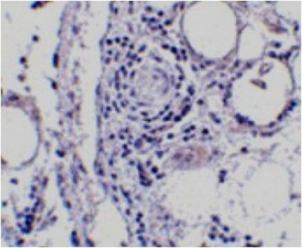
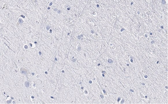
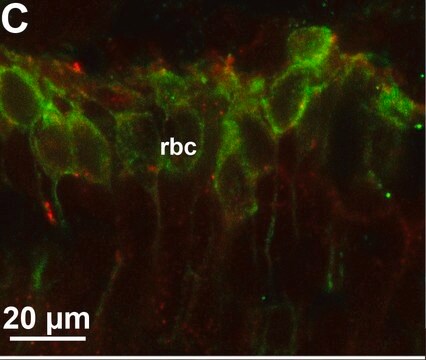
Immunohistochemistry Analysis: A 1:300 dilution of ABN14 antibody from a previous lot detected Brain lipid binding protein in mouse cerebellum tissue.
Research Category
Neuroscience
Research Sub Category
Neuronal & Glial Markers

Quality

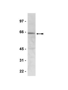
Evaluated by Western Blot in P1 mouse brain tissue lysate.

Western Blot Analysis: 0.01 µg/ml of this antibody detected Brain lipid-binding protein in 10 µg of P1 mouse brain tissue lysate.

Target description

~ 15 kDa

Linkage

Replaces: AB9558

Physical form

Affinity purified
Purified rabbit polyclonal in buffer containing 0.1 M Tris-Glycine (pH 7.4, 150 mM NaCl) with 0.05% sodium azide.

Storage and Stability

Stable for 1 year at 2-8°C from date of receipt.

Analysis Note

Control
P1 mouse brain tissue lysate

Other Notes

Concentration: Please refer to the Certificate of Analysis for the lot-specific concentration.

Disclaimer

Unless otherwise stated in our catalog or other company documentation accompanying the product(s), our products are intended for research use only and are not to be used for any other purpose, which includes but is not limited to, unauthorized commercial uses, in vitro diagnostic uses, ex vivo or in vivo therapeutic uses or any type of consumption or application to humans or animals.

Not finding the right product?  

Try our Product Selector Tool.

Storage Class

12 - Non Combustible Liquids

wgk_germany

WGK 1

flash_point_f

Not applicable

flash_point_c

Not applicable


Certificates of Analysis (COA)

Search for Certificates of Analysis (COA) by entering the products Lot/Batch Number. Lot and Batch Numbers can be found on a product’s label following the words ‘Lot’ or ‘Batch’.

Already Own This Product?

Find documentation for the products that you have recently purchased in the Document Library.

Visit the Document Library

Constance A Rich et al.
Glia, 66(12), 2617-2631 (2018-09-27)
We and others previously showed that in mouse embryos lacking the transcription factor Sox10, olfactory ensheathing cell (OEC) differentiation is disrupted, resulting in defective olfactory axon targeting and fewer gonadotropin-releasing hormone (GnRH) neurons entering the embryonic forebrain. The underlying mechanisms
Vivek K Bajpai et al.
Stem cells (Dayton, Ohio), 35(5), 1402-1415 (2017-02-01)
During development, neural crest (NC) cells are induced by signaling events at the neural plate border of all vertebrate embryos. Initially arising within the central nervous system, NC cells subsequently undergo an epithelial to mesenchymal transition to migrate into the
A mouse model of human primitive neuroectodermal tumors resulting from microenvironmentally-driven malignant transformation of orthotopically transplanted radial glial cells.
Malchenko, S; Sredni, ST; Hashimoto, H; Kasai, A; Nagayasu, K; Xie, J; Margaryan et al.
Testing null
Sara Anna Bonini et al.
Cerebral cortex (New York, N.Y. : 1991), 26(6), 2832-2849 (2016-03-08)
Alterations in genes that regulate neurodevelopment can lead to cortical malformations, resulting in malfunction during postnatal life. The NF-κB pathway has a key role during neurodevelopment by regulating the maintenance of the neural progenitor cell pool and inhibiting neuronal differentiation.
Mayumi Okamoto et al.
Nature communications, 7, 11349-11349 (2016-04-21)
During cerebral development, many types of neurons are sequentially generated by self-renewing progenitor cells called apical progenitors (APs). Temporal changes in AP identity are thought to be responsible for neuronal diversity; however, the mechanisms underlying such changes remain largely unknown.

Our team of scientists has experience in all areas of research including Life Science, Material Science, Chemical Synthesis, Chromatography, Analytical and many others.

Contact Technical Service