Skip to Content
MilliporeSigma
All Photos(1)

Key Documents

267A-1

Sigma-Aldrich

IgA Rabbit Polyclonal Antibody

Sign Into View Organizational & Contract Pricing


About This Item

UNSPSC Code:
12352203
NACRES:
NA.41

biological source

rabbit

Quality Level

100
500

conjugate

unconjugated

antibody form

Ig fraction of antiserum

antibody product type

primary antibodies

clone

polyclonal

description

For In Vitro Diagnostic Use in Select Regions (See Chart)

form

buffered aqueous solution

species reactivity

human

packaging

vial of 0.1 mL concentrate (267A-14)
vial of 0.5 mL concentrate (267A-15)
bottle of 1.0 mL predilute (267A-17)
vial of 1.0 mL concentrate (267A-16)
bottle of 7.0 mL predilute (267A-18)

manufacturer/tradename

Cell Marque

technique(s)

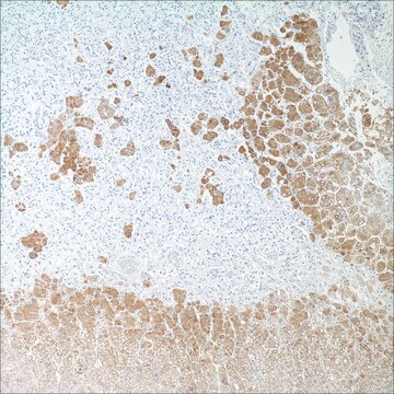
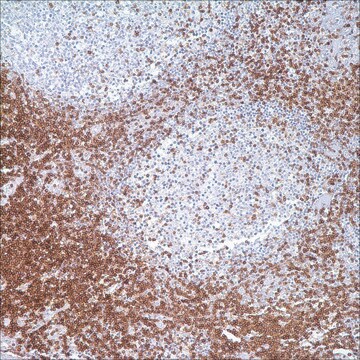
immunohistochemistry (formalin-fixed, paraffin-embedded sections): 1:100-1:500

control

tonsil

shipped in

wet ice

storage temp.

2-8°C

visualization

cytoplasmic

General description

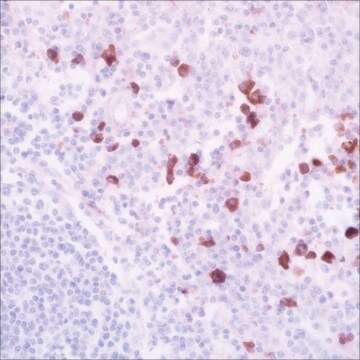
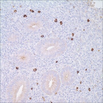
Anti-IgA antibody reacts with surface immunoglobulin IgA alpha chains. It is useful when identifying leukemias, plasmacytomas, and B-cell lineage derived Hodgkin′s lymphomas. Due to the restricted expression of heavy and light chains in these diseases, demonstration of B-cell lymphoma/plasmacytoma is aided with this antibody.

Quality


IVD

IVD

IVD

RUO

Linkage

IgA (polyclonal) Positive Control Slides, Product No. 267S, are available for immunohistochemistry (formalin-fixed, paraffin-embedded sections).

Physical form

Solution in Tris Buffer, pH 7.3-7.7, with 1% BSA and <0.1% Sodium Azide

Preparation Note

Download the IFU specific to your product lot and formatNote: This requires a keycode which can be found on your packaging or product label.

Other Notes

For Technical Service please contact: 800-665-7284 or email: service@cellmarque.com

Legal Information

Cell Marque is a trademark of Merck KGaA, Darmstadt, Germany

Not finding the right product?  

Try our Product Selector Tool.


Certificates of Analysis (COA)

Search for Certificates of Analysis (COA) by entering the products Lot/Batch Number. Lot and Batch Numbers can be found on a product’s label following the words ‘Lot’ or ‘Batch’.

Already Own This Product?

Find documentation for the products that you have recently purchased in the Document Library.

Visit the Document Library

Manual of Diagnostic Antibodies for Immunohistology, 217-219 (1999)
Tissue section immunologic methods in lymphomas
Warnake, R., et al.
Diagnostic Immunohistochemistry (Masson Publishing), 203-221 (1981)
Diffuse polyclonal B-cell lymphoma during primary infection with Epstein-Barr virus.
J E Robinson et al.
The New England journal of medicine, 302(23), 1293-1297 (1980-06-05)
C R Taylor
Archives of pathology & laboratory medicine, 102(3), 113-121 (1978-03-01)
Immunoperoxidase methods have much in common with established immunofluorescence procedures. Both have the potential for specific demonstration of cell and tissue antigens, with similar limitations demanding rigorous control of specificity. In any study the choice of an immunofluorescence method or
A Arnold et al.
The New England journal of medicine, 309(26), 1593-1599 (1983-12-29)
Immunoglobulin genes in their germ-line form are separated DNA subsegments that must be joined by means of recombinations during B-cell development. Individual immunoglobulin-gene rearrangements are specific for a given B cell and its progeny. We show that the detection of

Our team of scientists has experience in all areas of research including Life Science, Material Science, Chemical Synthesis, Chromatography, Analytical and many others.

Contact Technical Service