Skip to Content
MilliporeSigma
All Photos(1)

Key Documents

363M-1

Sigma-Aldrich

PAX-8 (MRQ-50) Mouse Monoclonal Antibody

Sign Into View Organizational & Contract Pricing


About This Item

UNSPSC Code:
12352203
NACRES:
NA.41

biological source

mouse

Quality Level

100
500

conjugate

unconjugated

antibody form

culture supernatant

antibody product type

primary antibodies

clone

MRQ-50, monoclonal

description

For In Vitro Diagnostic Use in Select Regions (See Chart)

form

buffered aqueous solution

species reactivity

human

packaging

vial of 0.1 mL concentrate (363M-14)
vial of 0.5 mL concentrate (363M-15)
bottle of 1.0 mL predilute (363M-17)
vial of 1.0 mL concentrate (363M-16)
bottle of 7.0 mL predilute (363M-18)

manufacturer/tradename

Cell Marque

technique(s)

immunohistochemistry: 1:50-1:200

isotype

IgG

shipped in

wet ice

storage temp.

2-8°C

Gene Information

human ... PAX8(7849)

General description

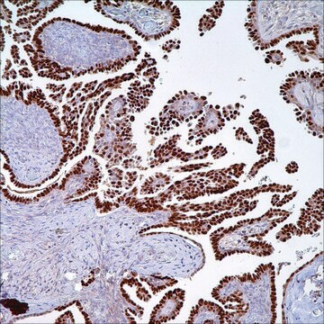
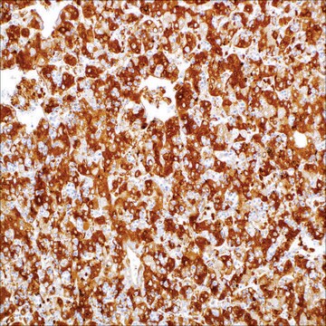
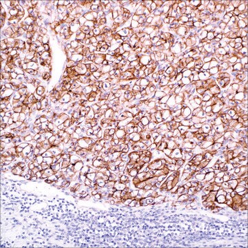
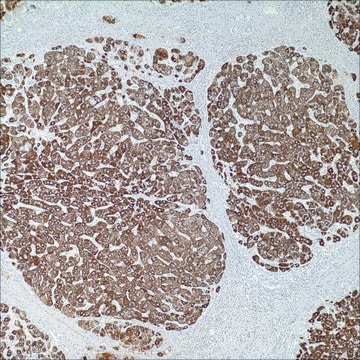
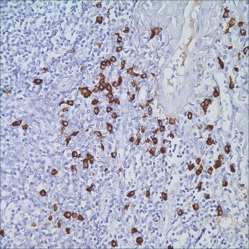
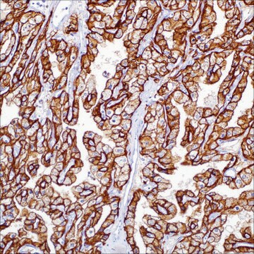
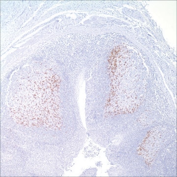
PAX-8 is a transcription factor expressed during embryonic development of Müllerian organs, kidneys, and thyroid, with continued expression in some epithelial cell types of these mature tissues.PAX-8 is a useful marker for neoplasms of gynecologic renal and thyroidorigin.PAX-8 (MRQ-50) antibody targets the N-terminus of the PAX-8 molecule. This section of the protein is shared with other PAX proteins such as PAX-5 and PAX-6; thus, some cross-reactivity with these proteins may be observed as evidenced by the labeling of selected lymphocytes, and neuroendocrine cells.

Quality


IVD

IVD

IVD

RUO

Physical form

Solution in Tris Buffer, pH 7.3-7.7, with 1% BSA and <0.1% Sodium Azide

Preparation Note

Download the IFU specific to your product lot and formatNote: This requires a keycode which can be found on your packaging or product label.

Other Notes

For Technical Service please contact: 800-665-7284 or email: service@cellmarque.com

Legal Information

Cell Marque is a trademark of Merck KGaA, Darmstadt, Germany

Not finding the right product?  

Try our Product Selector Tool.


Certificates of Analysis (COA)

Search for Certificates of Analysis (COA) by entering the products Lot/Batch Number. Lot and Batch Numbers can be found on a product’s label following the words ‘Lot’ or ‘Batch’.

Already Own This Product?

Find documentation for the products that you have recently purchased in the Document Library.

Visit the Document Library

Meaghan L Barr et al.
Journal of clinical pathology, 68(1), 12-17 (2014-10-16)
Immunohistochemical stains have greatly improved the diagnostic accuracy of renal cell carcinoma (RCC) for primary and distant tumours. We evaluate a marker that has recently been incorporated in clinical practice, PAX-8, in primary and metastatic RCCs. Two distinct tissue microarrays
Inigo Espinosa et al.
International journal of gynecological pathology : official journal of the International Society of Gynecological Pathologists, 34(3), 257-265 (2015-04-07)
Breast carcinomas rarely metastasize to the ovary and are even more rarely present clinically as primary ovarian tumors. However, patients with breast cancer not infrequently develop independent primary ovarian carcinomas. In these cases, distinction between independent primaries and metastatic tumors
Mark J Mentrikoski et al.
Applied immunohistochemistry & molecular morphology : AIMM, 22(9), 635-641 (2014-10-04)
Clear-cell renal cell carcinoma (CC-RCC) is the most common primary kidney malignancy, yet this morphology is not unique to renal primary tumors, as clear-cell variants of numerous nonrenal carcinomas of varying lineages exist. Therefore, because of CC-RCC's ability to metastasize
Ping Zhang et al.
Pathology international, 56(5), 240-245 (2006-05-04)
Thyroid transcription factor 1 (TTF1), thyroid transcription factor 2 (TTF2) and paired box gene 8 (Pax8) are demonstrated to play a crucial role for the differentiation and organogenesis of thyroid follicular cells. Their roles in thyroid carcinogenesis are not very
Ankur R Sangoi et al.
The American journal of surgical pathology, 35(5), 678-686 (2011-04-15)
The diagnosis of metastatic clear cell renal cell carcinoma (CC-RCC) can be difficult because of its morphologic heterogeneity and the increasing use of small image-guided biopsies that yield scant diagnostic material. This is further complicated by the degree of morphologic

Our team of scientists has experience in all areas of research including Life Science, Material Science, Chemical Synthesis, Chromatography, Analytical and many others.

Contact Technical Service