Skip to Content
MilliporeSigma
All Photos(2)

Documents

C8273

Sigma-Aldrich

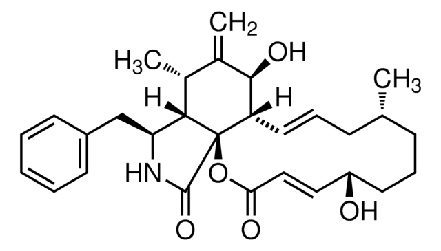
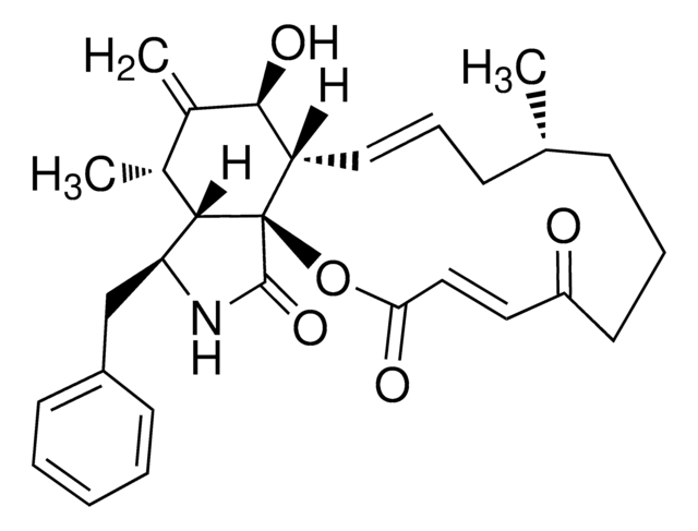
Cytochalasin D

from Zygosporium mansonii, ≥98% (TLC and HPLC), powder

Sign Into View Organizational & Contract Pricing

Synonym(s):
Lygosporin A, Zygosporin A
Empirical Formula (Hill Notation):
C30H37NO6
CAS Number:
Molecular Weight:
507.62
Beilstein/REAXYS Number:
1632828
EC Number:
MDL number:
UNSPSC Code:
12352200
PubChem Substance ID:
NACRES:
NA.77

biological source

Zygosporium mansonii

Quality Level

assay

≥98% (TLC and HPLC)

form

powder

solubility

DMSO: soluble
ethanol: soluble

antibiotic activity spectrum

fungi

mode of action

DNA synthesis | interferes

storage temp.

−20°C

SMILES string

[H][C@@]12[C@H](C)C(=C)[C@@H](O)[C@@H]3\C=C\C[C@H](C)C(=O)[C@](C)(O)\C=C\[C@@H](OC(C)=O)[C@@]13C(=O)N[C@H]2Cc4ccccc4

InChI

1S/C30H37NO6/c1-17-10-9-13-22-26(33)19(3)18(2)25-23(16-21-11-7-6-8-12-21)31-28(35)30(22,25)24(37-20(4)32)14-15-29(5,36)27(17)34/h6-9,11-15,17-18,22-26,33,36H,3,10,16H2,1-2,4-5H3,(H,31,35)/b13-9+,15-14+/t17-,18+,22-,23-,24+,25-,26+,29+,30+/m0/s1

InChI key

SDZRWUKZFQQKKV-JHADDHBZSA-N

Looking for similar products? Visit Product Comparison Guide

General description

Cytochalasin D (CytoD) is commonly used to inhibit actin dynamics and study its role in cellular processes. This fungal-derived small organic compound acts as an inhibitor of the interaction between G-actin and cofilin by binding to G-actin. Additionally, CytoD inhibits the binding of cofilin to F-actin and reduces the rate of actin polymerization and depolymerization in living cells.
Cytochalasin D is a cell-permeable fungal toxin. It disrupts actin microfilaments and activates the p53-dependent pathways causing arrest of the cell cycle at the G1-S transition. Cytochalasin D inhibits smooth muscle contraction and insulin-stimulated but not basal transport of glucose.

Application

Cytochalasin D has been used:
  • to treat H9c2 cells to study the relationship between cell stiffness and the F-actin cytoskeleton
  • as inhibitor of receptor-mediated endocytosis to treat oligodendrocyte precursor cells (OPCs) to investigate the effect of inhibiting specific extracellular vesicle (EV) uptake pathways
  • in in vitro embryo culture to artificially activate oocytes

Biochem/physiol Actions

Potent inhibitor of actin polymerization; disrupts actin microfilaments; activates the p53-dependent pathways; inhibits smooth muscle contraction; inhibits insulin-stimulated glucose transport.

related product

pictograms

Skull and crossbones

signalword

Danger

hcodes

Hazard Classifications

Acute Tox. 2 Oral

wgk_germany

WGK 3

ppe

Eyeshields, Faceshields, Gloves, type P3 (EN 143) respirator cartridges


Certificates of Analysis (COA)

Search for Certificates of Analysis (COA) by entering the products Lot/Batch Number. Lot and Batch Numbers can be found on a product’s label following the words ‘Lot’ or ‘Batch’.

Already Own This Product?

Find documentation for the products that you have recently purchased in the Document Library.

Visit the Document Library

Customers Also Viewed

Slide 1 of 7

1 of 7

Advances of xenogeneic ovarian extracellular matrix hydrogels for in vitro follicle development and oocyte maturation
Frances-Herrero E, et al.
Biomaterials advances (2023)
Hyperglycemia and hyperlipidemia can induce morphophysiological changes in rat cardiac cell line
Varela R, et al.
Biochemistry and Biophysics Reports (2021)
Extracellular vesicle-associated cholesterol supports the regenerative functions of macrophages in the brain
Vanherle S, et al.
Journal of Extracellular Vesicles (2023)
Cytochalasin D acts as an inhibitor of the actin-cofilin interaction
Shoji K, et al.
Biochemical and Biophysical Research Communications (2012)
Jennifer S N Verhoekx et al.
Journal of orthopaedic research : official publication of the Orthopaedic Research Society, 31(2), 328-334 (2012-09-19)
Matrix metalloproteinases (MMPs) are expressed in Dupuytren's contracture and play a role in matrix remodeling. We tested the role of tension on contractility and MMP expression in Dupuytren's nodule and cord cells. Cells were subjected to pre-determined loading patterns of

Articles

High titer lentiviral particles including beta-actin, alpha-tubulin and vimentin used for live cell analysis of cytoskeleton structure proteins.

Our team of scientists has experience in all areas of research including Life Science, Material Science, Chemical Synthesis, Chromatography, Analytical and many others.

Contact Technical Service