おすすめの製品
product name
Anti-Choline Acetyltransferase Antibody, clone 1E6, ascites fluid, clone 1E6, Chemicon®
由来生物
mouse
品質水準
抗体製品の状態
ascites fluid
抗体製品タイプ
primary antibodies
クローン
1E6, monoclonal
化学種の反応性
human, rat, monkey
メーカー/製品名
Chemicon®
テクニック
immunohistochemistry: suitable
アイソタイプ
IgG1
NCBIアクセッション番号
UniProtアクセッション番号
輸送温度
dry ice
ターゲットの翻訳後修飾
unmodified
遺伝子情報
human ... CHAT(1103)
rat ... Chat(290567)
rhesus monkey ... Chat(709977)
特異性
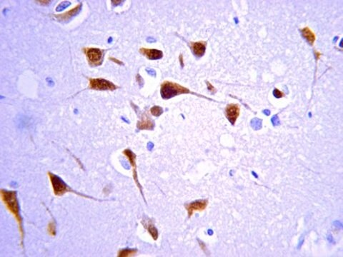
脳および脊髄(CNS)のコリン作動性ニューロンを認識します。
免疫原
ラット脳から精製されたコリンアセチルトランスフェラーゼ。
アプリケーション
Immunohistochemistry: 1:100-1:250. See immunohistochmistry procedure below.
Optimal working dilutions must be determined by the end user.
IMMUNOHISTOCHEMISTRY PROCEDURE (PAP TECHNIQUE) FOR MAB305, MONOCLONAL ANTIBODY TO CHOLINE ACETYLTRANSFERASE
I) Perfusion & Sectioning Procedure
1. Perfuse through the heart with a fixative solution containing 4% paraformaldehyde in 0.12 M phosphate buffer (pH 7.3) for light microscopy (LM), and additionally, 0.1% gluteraldehyde and .002% CaCl2 for electron microscopy (EM).
2. Remove brain and postfix 2-18 hours at 4°C in 4% paraformaldehyde in 0.12 M phosphate buffer.
3. After brain is blocked for sectioning, wash in several changes of buffer for 2-3 hours.
4. Specimens for EM are sectioned on a Vibratome (50 μm) and rinsed in buffer, those for LM should be cryoprotected in 30% sucrose in buffer.
5. After freezing with dry ice, 30-40 μm thick sections of LM specimens are cut on a cryostat.
6. Sections are rinsed, and then stored in phosphate buffer containing 0.1% sodium azide.
II) Staining Procedure
Tissue is processed as freely-floating sections in continuously agitated solutions. All incubations are performed at room temperature unless otherwise stated.
1.a. For localizing ChAT-positive somata and dendrites:
Sections are washed in 0.1 M Tris-buffered saline (TBS; containing 1.4% NaCl, pH 7.3) only. No detergent or enzyme pretreatment is used.
b. For localizing ChAT-positive terminal-like structures:
Incubate sections in TBS (pH 8.1) for 5 minutes at 37°C. Transfer sections to TBS (pH 8.1) containing pronase (1.2 μg/mL) for 1 1/2-2 minutes at 37°C, followed by several ice cold buffer washes for a total of 5 minutes. The concentration of pronase and incubation time of the digestion should be evaluated for each region examined.
c. For localizing ChAT immunoreactivity and subsequently counterstaining the sections:
Incubation in TBS containing 0.1%-0.8% Triton X-100 for 15 minutes may increase the tissue penetration of the immunoreagents, but it also raises the background staining.
2. Incubate sections in normal goat serum (3-5%) for one hour. The working solutions of all antisera should also contain similarly diluted normal goat serum.
3. Incubate in anti-ChAT monoclonal antibody solution (Suggested working dilution 1:250, final working dilution must be determined by end user) for 2 hours at room temperature and then for an additional 6-18 hours at 4°C.
4. Incubate with second antibody (i.e. Goat anti-Mouse IgG, Cat. No.: AP124, dilution 1:50-100) for 1-2 hours.
5. Incubate with diluted PAP complex (i.e. Mouse PAP, Cat No.: PAP14, conc. 25-50 μg/mL) for one hour.
6. After rinsing in buffer, the second antibody and PAP steps are repeated for 40 minutes to 1 hour each in order to amplify staining intensity, particularly of small ChAT-containing structures.
7. React for 15 minutes with 0.06% 3,3′-diaminobenzidine×4 HCl (DAB; diluted in phosphate buffered saline, pH 7.3) and 0.006% H2O2.
8. Specimens for routine LM are postfixed for 1 minutes in 0.005% OsO4 (osmium tetraoxide), and then mounted, dehydrated and coverslipped. Selected regions blocked for EM are postfixed in 2% OsO4 for 1 hour, en bloc stained with uranyl acetate, and flat-embedded in Epon-Araldite resin.
この抗コリンアセチルトランスフェラーゼ抗体、クローン1E6を用いたコリンアセチルトランスフェラーゼの検出は、IHでの使用が検証されています。
研究のカテゴリ
神経科学
神経科学
研究サブカテゴリー
神経伝達物質および受容体
神経細胞およびグリアマーカー
神経伝達物質および受容体
神経細胞およびグリアマーカー
物理的形状
未精製
防腐剤を含まない腹水。
保管および安定性
-20°Cで出荷日から1年間保存できます。凍結融解を繰り返さないために、分注してください。製品を最大限回収するために、融解後のキャップを外す前に元のバイアルを遠心分離します。
アナリシスノート
コントロール
脳組織
脳組織
その他情報
濃度:ロットに固有の濃度につきましては試験成績書をご参照ください。
法的情報
CHEMICON is a registered trademark of Merck KGaA, Darmstadt, Germany
免責事項
メルクのカタログまたは製品に添付されたメルクのその他の文書に記載されていない場合、メルクの製品は研究用途のみを目的としているため、他のいかなる目的にも使用することはできません。このような目的としては、未承認の商業用途、in vitroの診断用途、ex vivoあるいはin vivoの治療用途、またはヒトあるいは動物へのあらゆる種類の消費あるいは適用などがありますが、これらに限定されません。
適切な製品が見つかりませんか。
製品選択ツール.をお試しください
保管分類コード
10 - Combustible liquids
WGK
WGK 1
引火点(°F)
Not applicable
引火点(℃)
Not applicable
適用法令
試験研究用途を考慮した関連法令を主に挙げております。化学物質以外については、一部の情報のみ提供しています。 製品を安全かつ合法的に使用することは、使用者の義務です。最新情報により修正される場合があります。WEBの反映には時間を要することがあるため、適宜SDSをご参照ください。
Jan Code
MAB305:
試験成績書(COA)
製品のロット番号・バッチ番号を入力して、試験成績書(COA) を検索できます。ロット番号・バッチ番号は、製品ラベルに「Lot」または「Batch」に続いて記載されています。
Characterization of cognitive deficits in rats overexpressing human alpha-synuclein in the ventral tegmental area and medial septum using recombinant adeno-associated viral vectors.
Hall, H; Jewett, M; Landeck, N; Nilsson, N; Schagerlof, U; Leanza, G; Kirik, D
Testing null
Naitao Wang et al.
Stem cell reports, 6(5), 668-678 (2016-05-12)
Regulation of prostate epithelial progenitor cells is important in prostate development and prostate diseases. Our previous study demonstrated a function of autocrine cholinergic signaling (ACS) in promoting prostate cancer growth and castration resistance. However, whether or not such ACS also
Glucocorticoid receptors in the locus coeruleus mediate sleep disorders caused by repeated corticosterone treatment.
Wang, ZJ; Zhang, XQ; Cui, XY; Cui, SY; Yu, B; Sheng, ZF; Li, SJ; Cao, Q; Huang, YL; Xu et al.
Scientific Reports null
Samantha R Eck et al.
Biological psychiatry, 88(7), 566-575 (2020-07-01)
Stress exacerbates symptoms of schizophrenia and attention-deficit/hyperactivity disorder, which are characterized by impairments in sustained attention. Yet how stress regulates attention remains largely unexplored. We investigated whether a 6-day variable stressor altered sustained attention and the cholinergic attention system in
C Alex Goddard et al.
Journal of neurophysiology, 98(6), 3486-3493 (2007-09-28)
Cholinergic neurons in the parabigeminal nucleus of the rat midbrain were studied in an acute slice preparation. Spontaneous, regular action potentials were observed both with cell-attached patch recordings as well as with whole cell current-clamp recordings. The spontaneous activity of
ライフサイエンス、有機合成、材料科学、クロマトグラフィー、分析など、あらゆる分野の研究に経験のあるメンバーがおります。.
製品に関するお問い合わせはこちら(テクニカルサービス)