추천 제품
일반 설명
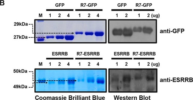
The Green Fluorescent Protein (GFP) from the jellyfish Aequorea victoria is used as a fluorescent indicator for monitoring gene expression in a variety of cellular systems, including living organisms and fixed tissues. Unlike other bioluminescent reporters, GFP fluoresces in the absence of substrates, cofactors, or other intrinsic or extrinsic proteins. Purified GFP is a 27 kDa monomer consisting of 238 amino acids and emits green light (emission maximum at 509 nm) when excited with blue or UV light.
특이성
AB16901 is made against highly-purified native GFP from Aequorea victoria. It is reactive with GFP from both native and recombinant sources.
면역원
A recombinant green fluorescent protein (GFP) containing a 6-his tag, was highly purified by affinity chromatography on Nickel column followed by chromatography on a P-100 gel filtration column. The resulting product was present as only a singl
애플리케이션
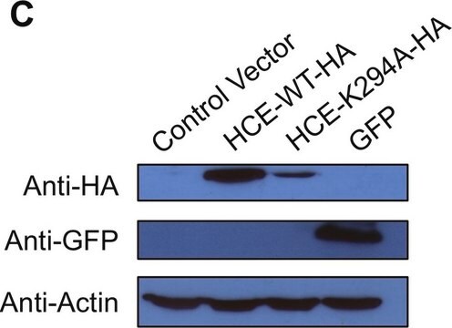
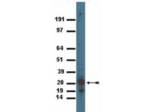
Immunoblotting: AB16901 detects a band of the proper molecular weight (30kDa) in lysates from E.coli expressing GFP but not in E.coli that do not express or in lysates prepared from E.coli containing the GFP-expressing plasmid but not induced, detect GFP in lysates from COS or primary neurons or fibroblast cells transfected with plasmids directing expression GFP or a tau-GFP fusion protein. Did not detect any signal when 50μg of protein from a rat brain lysate (negative control) was fractionated on the SDS-PAGE. Titers were 10-20,000 when detected with an anti-chicken horseradish peroxidase-conjugated secondary by ECL or by a colorimetric assay using DAB.
Immunocytochemistry: GFP can be detected in cells that are transfected with a plasmid directing the expression of either GFP or a GFP-Fusion protein. Cells that do not express GFP exhibit no detectable staining. AB binding was determined using HRP conjugated-anti chicken secondary Abs and visualized using DAB. The anti-GFP Abs were used at 1-2,500-5,000 fold dilution. Cells were fixed using 4% formaldehyde in PBS (pH7.4). For immunocytochemistry, the cultures were fixed in 4% formaldehyde in PBS pH7.4, for 20 minutes at room temperature. The cells are permeabilized with 0.2% Triton X-100 and 2% serum (corresponding to the aminal in which the secondary antibody was raised) in PBS for 20 minutes at room temperature. Cell cultures are then incubated with primary antisera in PBS containing 2% serum (as above), at 4°C, overnight. Antibody binding is detected using an avidin/biotin/peroxidase detection system. Cultures were washed four times (10 minutes each) between steps, and reaction product is developed with a 0.05% diaminobenzidine / 0.02% hydrogen peroxide solution on ice for 8-10 minutes. AB16901 has been used to detect GFP in primary neurons and zebrafish fibroblasts.
Immunoprecipitation: 5μg antibody per no more than 500μl of cell lysate. It is important to use a rabbit anti-chicken secondary for capture or anti-chicken beads as Chicken IgG will not react with protein A or protein G.
Optimal working dilutions must be determined by the end user.
Research Category
Epitope Tags & General Use
Epitope Tags & General Use
Research Sub Category
Epitope Tags
Epitope Tags
This Anti-Green Fluorescent Protein Antibody is validated for use in IC, IP, WB for the detection of Green Fluorescent Protein.
표적 설명
MW for GFP is 27 kDa Dependent upon the molecular weight of the fusion protein being detected.
물리적 형태
Ammonium Sulfate Precipitation
Format: Purified
Purified chicken immunoglobulin in TBS, pH 7.5 with 0.02% sodium azide.
저장 및 안정성
The undiluted antibody solution is stable for approximately 6 months stored between 2 and 8°C.
During shipment, small volumes of product will occasionally become entrapped in the seal of the product vial. For products with volumes of 200μL or less, we recommend gently tapping the vial on a hard surface or briefly centrifuging the vial in a tabletop centrifuge to dislodge any liquid in the container′s cap.
During shipment, small volumes of product will occasionally become entrapped in the seal of the product vial. For products with volumes of 200μL or less, we recommend gently tapping the vial on a hard surface or briefly centrifuging the vial in a tabletop centrifuge to dislodge any liquid in the container′s cap.
분석 메모
Control
GFP fusion protein expressed in cells
GFP fusion protein expressed in cells
기타 정보
Concentration: Please refer to the Certificate of Analysis for the lot-specific concentration.
법적 정보
CHEMICON is a registered trademark of Merck KGaA, Darmstadt, Germany
면책조항
Unless otherwise stated in our catalog or other company documentation accompanying the product(s), our products are intended for research use only and are not to be used for any other purpose, which includes but is not limited to, unauthorized commercial uses, in vitro diagnostic uses, ex vivo or in vivo therapeutic uses or any type of consumption or application to humans or animals.
적합한 제품을 찾을 수 없으신가요?
당사의 제품 선택기 도구.을(를) 시도해 보세요.
Storage Class Code
12 - Non Combustible Liquids
WGK
nwg
Flash Point (°F)
Not applicable
Flash Point (°C)
Not applicable
시험 성적서(COA)
제품의 로트/배치 번호를 입력하여 시험 성적서(COA)을 검색하십시오. 로트 및 배치 번호는 제품 라벨에 있는 ‘로트’ 또는 ‘배치’라는 용어 뒤에서 찾을 수 있습니다.
이미 열람한 고객
T Fukushige et al.
Proceedings of the National Academy of Sciences of the United States of America, 96(21), 11883-11888 (1999-10-16)
In analyzing the transcriptional networks that regulate development, one ideally would like to determine whether a particular transcription factor binds directly to a candidate target promoter inside the living embryo. Properties of the Caenorhabditis elegans elt-2 gene, which encodes a
Eunhee Kim et al.
Journal of cerebral blood flow and metabolism : official journal of the International Society of Cerebral Blood Flow and Metabolism, 34(8), 1411-1419 (2014-05-29)
Monocytes/macrophages (MMs), mononuclear phagocytes, have been implicated in stroke-induced inflammation and injury. However, the presence of pro-inflammatory Ly-6C(high) and antiinflammatory Ly-6C(low) monocyte subsets raises uncertainty regarding their role in stroke pathologic assessment. With recent identification of the spleen as an
Temporal characterization of homology-independent centromere coupling in meiotic prophase.
Obeso, D; Dawson, DS
Testing null
Robert Andrews et al.
PloS one, 2(6), e493-e493 (2007-06-07)
Endocytosis is involved in the regulation of many cellular events, including signalling, cell migration, and cell polarity. To begin to investigate roles for endocytosis in early C. elegans development, we examined the distribution and dynamics of early endosomes (EEs) in
Chenshu Liu et al.
The Journal of cell biology, 213(4), 415-424 (2016-05-18)
Centromeres of higher eukaryotes are epigenetically defined by centromere protein A (CENP-A), a centromere-specific histone H3 variant. The incorporation of new CENP-A into centromeres to maintain the epigenetic marker after genome replication in S phase occurs in G1 phase; however
자사의 과학자팀은 생명 과학, 재료 과학, 화학 합성, 크로마토그래피, 분석 및 기타 많은 영역을 포함한 모든 과학 분야에 경험이 있습니다..
고객지원팀으로 연락바랍니다.
