
추천 제품


생물학적 소스
mouse
Quality Level
결합
unconjugated
항체 형태
purified immunoglobulin
항체 생산 유형
primary antibodies
클론
13.1, monoclonal
7.1, monoclonal
분석
>90% (HPLC)
양식
lyophilized
포장
pkg of 200 μg
제조업체/상표
Roche
동형
IgG1κ
저장 온도
2-8°C
일반 설명
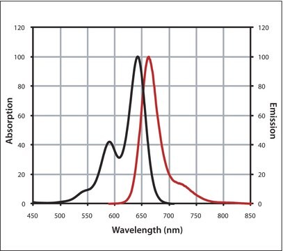
Green Fluorescent Protein (GFP) is a spontaneously fluorescent 27kDa protein originally isolated from the jellyfish Aequorea victoria. The molecular cloning of the GFP gene and its subsequent expression in heterologous systems has established GFP as a valuable reporter molecule for in vivo visualization of gene expression events in a wide variety of cell types and organisms. Since, GFP requires no additional substrates or cofactors, GFP′s green fluorescence can be easily detected using blue or UV light after expression in either prokaryotic or eukaryotic cells. In addition, several mutant forms of GFP with unique spectral properties (e.g., enhanced fluorescence signal and shifts in excitation and emission spectra) have been reported.
Mixture of two high-affinity monoclonal antibodies selected for their performance in detection of GFP and GFP fusion proteins.
특이성
Subtype: Both clones are Mouse IgG1κ
애플리케이션
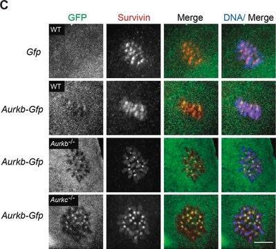
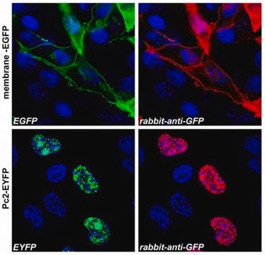
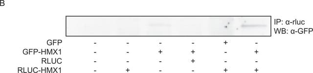
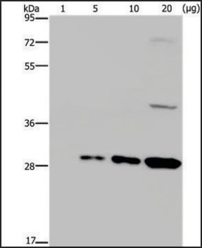
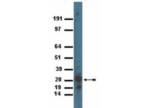
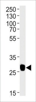
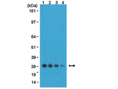
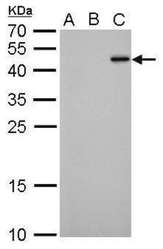
Monoclonal antibody for detection of both wild-type and mutant forms of GFP or GFP fusions using:
- Immunoprecipitation
- Western blots
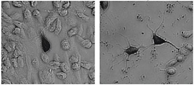
- Immunostaining
특징 및 장점
Contents
Mixture of two monoclonal antibodies, supplied as a white lyophilizatecontaining 200μg of total Anti-GFP IgG.
Anti-GFP is a mixture of two clones (7.1 and 13.1).
Mixture of two monoclonal antibodies, supplied as a white lyophilizatecontaining 200μg of total Anti-GFP IgG.
Anti-GFP is a mixture of two clones (7.1 and 13.1).
품질
Anti-GFP is tested for functionality and purity relative to a reference standard to confirm the quality of each new reagent preparation.
Purity: Both Anti-GFP mouse monoclonal antibodies (Clones 7.1 and 13.1) are >95% pure as determined by SDS-PAGE and ion-exchange HPLC analyses.
Purity: Both Anti-GFP mouse monoclonal antibodies (Clones 7.1 and 13.1) are >95% pure as determined by SDS-PAGE and ion-exchange HPLC analyses.
제조 메모
Working concentration: Working concentration of antibody depends on application and substrate.
The following concentrations should be taken as a guideline:
Storage conditions (working solution): -15 to -25 °C
The following concentrations should be taken as a guideline:
- Western blot: 1:1000 dilution
- Immunoprecipitation: 2 to 10 μg
Storage conditions (working solution): -15 to -25 °C
재구성
Add 500 μl double distilled water to a final concentration of 0.4 mg/ml.
Rehydrate on ice for 30 minutes.
Rehydrate on ice for 30 minutes.
법적 정보
This product is sold under license from Columbia University. Rights to use this product are limited to research use only. No other rights are conveyed. Inquiry into the availability of a license to broader rights or the use of this product for commercial purposes should be directed to Columbia Innovation Enterprise, Columbia University, Engineering Terrace - Suite 363, new York, New York 10027.
적합한 제품을 찾을 수 없으신가요?
당사의 제품 선택기 도구.을(를) 시도해 보세요.
Storage Class Code
13 - Non Combustible Solids
WGK
WGK 1
Flash Point (°F)
does not flash
Flash Point (°C)
does not flash
이미 열람한 고객

Ward W.W, et al.
Photochemistry and Photobiology, 31, 611-615 (1980)
Chalfie M, et al.
Science, 263, 802-805 (1994)
Prasher D C, et al.
Gene, 111, 229-233 (1992)
Chi C Wong et al.
Blood, 118(16), 4305-4312 (2011-08-02)
Shwachman-Diamond syndrome (SDS), a recessive leukemia predisposition disorder characterized by bone marrow failure, exocrine pancreatic insufficiency, skeletal abnormalities and poor growth, is caused by mutations in the highly conserved SBDS gene. Here, we test the hypothesis that defective ribosome biogenesis
Crameri A, et al.
Nature Biotechnology, 14, 315-319 (1996)
자사의 과학자팀은 생명 과학, 재료 과학, 화학 합성, 크로마토그래피, 분석 및 기타 많은 영역을 포함한 모든 과학 분야에 경험이 있습니다..
고객지원팀으로 연락바랍니다.