
추천 제품


제품명
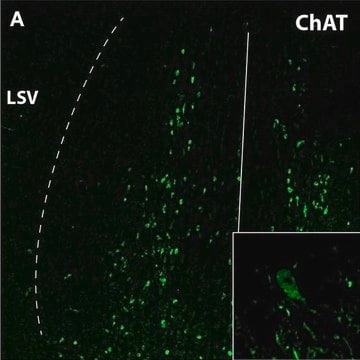
Anti-Choline Acetyltransferase Antibody, clone 1E6, ascites fluid, clone 1E6, Chemicon®
생물학적 소스
mouse
Quality Level
항체 형태
ascites fluid
항체 생산 유형
primary antibodies
클론
1E6, monoclonal
종 반응성
human, rat, monkey
제조업체/상표
Chemicon®
기술
immunohistochemistry: suitable
동형
IgG1
UniProt 수납 번호
배송 상태
dry ice
타겟 번역 후 변형
unmodified
유전자 정보
human ... CHAT(1103)
rat ... Chat(290567)
rhesus monkey ... Chat(709977)
특이성
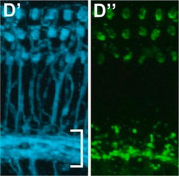
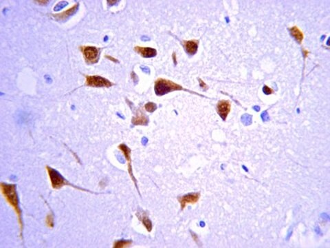
Recognizes cholinergic neurons in the brain and spinal cord (CNS).
면역원
Choline acetyltransferase purified from rat brain.
애플리케이션
Detect Choline Acetyltransferase using this Anti-Choline Acetyltransferase Antibody, clone 1E6 validated for use in IH.
Immunohistochemistry: 1:100-1:250. See immunohistochmistry procedure below.
Optimal working dilutions must be determined by the end user.
IMMUNOHISTOCHEMISTRY PROCEDURE (PAP TECHNIQUE) FOR MAB305, MONOCLONAL ANTIBODY TO CHOLINE ACETYLTRANSFERASE
I) Perfusion & Sectioning Procedure
1. Perfuse through the heart with a fixative solution containing 4% paraformaldehyde in 0.12 M phosphate buffer (pH 7.3) for light microscopy (LM), and additionally, 0.1% gluteraldehyde and .002% CaCl2 for electron microscopy (EM).
2. Remove brain and postfix 2-18 hours at 4°C in 4% paraformaldehyde in 0.12 M phosphate buffer.
3. After brain is blocked for sectioning, wash in several changes of buffer for 2-3 hours.
4. Specimens for EM are sectioned on a Vibratome (50 μm) and rinsed in buffer, those for LM should be cryoprotected in 30% sucrose in buffer.
5. After freezing with dry ice, 30-40 μm thick sections of LM specimens are cut on a cryostat.
6. Sections are rinsed, and then stored in phosphate buffer containing 0.1% sodium azide.
II) Staining Procedure
Tissue is processed as freely-floating sections in continuously agitated solutions. All incubations are performed at room temperature unless otherwise stated.
1.a. For localizing ChAT-positive somata and dendrites:
Sections are washed in 0.1 M Tris-buffered saline (TBS; containing 1.4% NaCl, pH 7.3) only. No detergent or enzyme pretreatment is used.
b. For localizing ChAT-positive terminal-like structures:
Incubate sections in TBS (pH 8.1) for 5 minutes at 37°C. Transfer sections to TBS (pH 8.1) containing pronase (1.2 μg/mL) for 1 1/2-2 minutes at 37°C, followed by several ice cold buffer washes for a total of 5 minutes. The concentration of pronase and incubation time of the digestion should be evaluated for each region examined.
c. For localizing ChAT immunoreactivity and subsequently counterstaining the sections:
Incubation in TBS containing 0.1%-0.8% Triton X-100 for 15 minutes may increase the tissue penetration of the immunoreagents, but it also raises the background staining.
2. Incubate sections in normal goat serum (3-5%) for one hour. The working solutions of all antisera should also contain similarly diluted normal goat serum.
3. Incubate in anti-ChAT monoclonal antibody solution (Suggested working dilution 1:250, final working dilution must be determined by end user) for 2 hours at room temperature and then for an additional 6-18 hours at 4°C.
4. Incubate with second antibody (i.e. Goat anti-Mouse IgG, Cat. No.: AP124, dilution 1:50-100) for 1-2 hours.
5. Incubate with diluted PAP complex (i.e. Mouse PAP, Cat No.: PAP14, conc. 25-50 μg/mL) for one hour.
6. After rinsing in buffer, the second antibody and PAP steps are repeated for 40 minutes to 1 hour each in order to amplify staining intensity, particularly of small ChAT-containing structures.
7. React for 15 minutes with 0.06% 3,3′-diaminobenzidine×4 HCl (DAB; diluted in phosphate buffered saline, pH 7.3) and 0.006% H2O2.
8. Specimens for routine LM are postfixed for 1 minutes in 0.005% OsO4 (osmium tetraoxide), and then mounted, dehydrated and coverslipped. Selected regions blocked for EM are postfixed in 2% OsO4 for 1 hour, en bloc stained with uranyl acetate, and flat-embedded in Epon-Araldite resin.
Research Category
Neuroscience
Neuroscience
Research Sub Category
Neurotransmitters & Receptors
Neuronal & Glial Markers
Neurotransmitters & Receptors
Neuronal & Glial Markers
물리적 형태
Ascites fluid containing no preservatives.
Unpurified
저장 및 안정성
Maintain for 1 year at -20°C from date of shipment. Aliquot to avoid repeated freezing and thawing. For maximum recovery of product, centrifuge the original vial after thawing and prior to removing the cap.
분석 메모
Control
Brain tissue
Brain tissue
기타 정보
Concentration: Please refer to the Certificate of Analysis for the lot-specific concentration.
법적 정보
CHEMICON is a registered trademark of Merck KGaA, Darmstadt, Germany
면책조항
Unless otherwise stated in our catalog or other company documentation accompanying the product(s), our products are intended for research use only and are not to be used for any other purpose, which includes but is not limited to, unauthorized commercial uses, in vitro diagnostic uses, ex vivo or in vivo therapeutic uses or any type of consumption or application to humans or animals.
적합한 제품을 찾을 수 없으신가요?
당사의 제품 선택기 도구.을(를) 시도해 보세요.
Storage Class Code
10 - Combustible liquids
WGK
WGK 1
Flash Point (°F)
Not applicable
Flash Point (°C)
Not applicable
시험 성적서(COA)
제품의 로트/배치 번호를 입력하여 시험 성적서(COA)을 검색하십시오. 로트 및 배치 번호는 제품 라벨에 있는 ‘로트’ 또는 ‘배치’라는 용어 뒤에서 찾을 수 있습니다.
Tamunotonye Omoluabi et al.
Alzheimer's & dementia (New York, N. Y.), 7(1), e12231-e12231 (2022-01-11)
The earliest abnormality associated with Alzheimer's disease (AD) is the presence of persistently phosphorylated pretangle tau in locus coeruleus (LC) neurons. LC neuron numbers and fiber density are positive predictors of cognition prior to death. Using an animal model of
Chondroitinase applied to peripheral nerve repair averts retrograde axonal regeneration.
Graham, JB; Neubauer, D; Xue, QS; Muir, D
Experimental neurology null
C Alex Goddard et al.
Journal of neurophysiology, 98(6), 3486-3493 (2007-09-28)
Cholinergic neurons in the parabigeminal nucleus of the rat midbrain were studied in an acute slice preparation. Spontaneous, regular action potentials were observed both with cell-attached patch recordings as well as with whole cell current-clamp recordings. The spontaneous activity of
Glucocorticoid receptors in the locus coeruleus mediate sleep disorders caused by repeated corticosterone treatment.
Wang, ZJ; Zhang, XQ; Cui, XY; Cui, SY; Yu, B; Sheng, ZF; Li, SJ; Cao, Q; Huang, YL; Xu et al.
Scientific Reports null
Naitao Wang et al.
Stem cell reports, 6(5), 668-678 (2016-05-12)
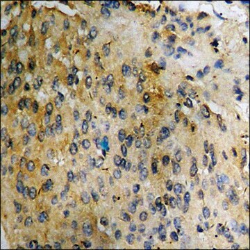
Regulation of prostate epithelial progenitor cells is important in prostate development and prostate diseases. Our previous study demonstrated a function of autocrine cholinergic signaling (ACS) in promoting prostate cancer growth and castration resistance. However, whether or not such ACS also
자사의 과학자팀은 생명 과학, 재료 과학, 화학 합성, 크로마토그래피, 분석 및 기타 많은 영역을 포함한 모든 과학 분야에 경험이 있습니다..
고객지원팀으로 연락바랍니다.