
06-735
Anti-Caspase 3 Antibody
Upstate®, from rabbit
Synonym(s):
Anti-CASP-3, Anti-Caspase-3
Sign Into View Organizational & Contract Pricing
All Photos(3)
About This Item
UNSPSC Code:
12352203
eCl@ss:
32160702
NACRES:
NA.41
Pricing and availability is not currently available.
Recommended Products


General description
Caspase-3 (UniProt: P42574; also known as EC:3.4.22.56, CASP-3, Apopain, Cysteine protease CPP32, CPP-32, Protein Yama, SREBP cleavage activity 1, SCA-1) is encoded by the CASP3 (also known as CPP32) gene (Gene ID: 836) in human. Cysteine-aspartic proteases or Caspases play essential roles in apoptosis, necrosis, and inflammation. Historically, caspases were numbered in the order in which they were identified. Caspase-3 is a heterotetrameric enzyme that consists of two anti-parallel arranged heterodimers, each one formed by a 17 kDa (p17) and a 12 kDa (p12) subunit. Caspase-3 is initially produced with a propeptide sequence (aa 1-9), the removal of which yields the 268 aa. caspase-3 proenzyme. Upon activation, the proenzyme is proteolytically cleaved first between Asp175-Ser176 to generate a p20 (aa 10-175) fragment and the p12 (aa 176-277) subunit. Further cleavage of the p20 fragment between Asp28-Ser29 produces the p17 (aa 29-175) subunit. The p17 and p12 subunits dimerize and forms the active caspase-3 enzyme. Caspase-3 has a strict requirement for an Asp residue at positions P1 and P4. It has a preferred cleavage sequence of Asp-Xaa-Xaa-Asp-|- with a hydrophobic amino-acid residue at P2 and a hydrophilic amino-acid residue at P3, although Val or Ala are also accepted at this position. Caspase-3 is involved in the activation cascade of caspases responsible for apoptosis execution. At the onset of apoptosis, it proteolytically cleaves poly(ADP-ribose) polymerase (PARP) at a Asp216-|-Gly217 bond. Caspase-3 mediates the proteolytic activation of caspases-6 and -7, while caspase-3 itself is processed and activated by caspase-8, -9, or -10.
Specificity
Recognizes full-length Caspase 3 (Yama/Apopain) and proteolytic fragments.
Immunogen
Human full-length Caspase 3 fusion protein containing a histidine-6 tag
Application
This Anti-Caspase 3 Antibody is validated for use in Immunihistochmistry and Western Blotting for the detection of Caspase 3.
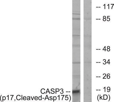
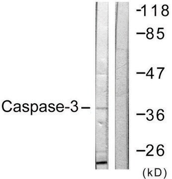
Western Blotting Analysis: 1μg/mL of this antibody detects Caspase-3 in A431 cell lysate.
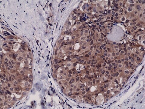
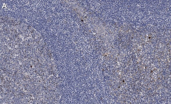
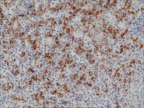
Immunohistochemistry (Paraffin) Analysis: A 1:250 dilution of this antibody detected Caspase-3 in Human tonsil tissue sections.
Immunohistochemistry (Paraffin) Analysis: A 1:250 dilution of this antibody detected Caspase-3 in Human tonsil tissue sections.
Quality
routinely evaluated by immunoblot on RIPA lysates from non-stimulated human A431 cells, mouse 3T3/A31 or rat PC12 cells
Target description
32 kDa
Linkage
Replaces: 04-1090; 04-439
Physical form
Format: Purified
Protein A purified IgG in of 0.1M Tris-glycine, pH 7.4, 0.15M NaCl,and 0.05% sodium azide.
Storage and Stability
Stable for 2 years at 2-8°C from date of shipment. For maximum recovery of product, centrifuge the original vial prior to removing the cap.
Analysis Note
Control
Positive Antigen Control: Catalog #12-301, non-stimulated A431 cell lysate. Add 2.5µL of 2-mercaptoethanol/100µL of lysate and boil for 5 minutes to reduce the preparation. Load 20µg of reduced lysate per lane for mingels.
Positive Antigen Control: Catalog #12-301, non-stimulated A431 cell lysate. Add 2.5µL of 2-mercaptoethanol/100µL of lysate and boil for 5 minutes to reduce the preparation. Load 20µg of reduced lysate per lane for mingels.
Legal Information
UPSTATE is a registered trademark of Merck KGaA, Darmstadt, Germany
Not finding the right product?
Try our Product Selector Tool.
recommended
Product No.
Description
Pricing
Storage Class
10 - Combustible liquids
wgk_germany
WGK 1
Certificates of Analysis (COA)
Search for Certificates of Analysis (COA) by entering the products Lot/Batch Number. Lot and Batch Numbers can be found on a product’s label following the words ‘Lot’ or ‘Batch’.
Already Own This Product?
Find documentation for the products that you have recently purchased in the Document Library.
Customers Also Viewed


Antiandrogen-induced cell death in LNCaP human prostate cancer cells.
Lee, EC; Zhan, P; Schallhom, R; Packman, K; Tenniswood, M
Cell Death and Differentiation null
A caspase cascade regulating developmental axon degeneration.
Simon, DJ; Weimer, RM; McLaughlin, T; Kallop, D; Stanger, K; Yang, J; O'Leary et al.
The Journal of Neuroscience null
Prisca Boisguérin et al.
Cardiovascular research, 116(3), 633-644 (2019-05-31)
Regulated cell death is a main contributor of myocardial ischaemia-reperfusion (IR) injury during acute myocardial infarction. In this context, targeting apoptosis could be a potent therapeutical strategy. In a previous study, we showed that DAXX (death-associated protein) was essential for
M Leist et al.
Biochemical and biophysical research communications, 258(1), 215-221 (1999-05-01)
The endogenous mediator nitric oxide (NO) blocked apoptosis of Jurkat cells elicited by staurosporine, anti-CD95 or chemotherapeutics, and switched death to necrosis. The switch in the mode of cell death was dependent on the ATP loss elicited by NO. This
Chiang-Ting Chien et al.
Journal of the American Society of Nephrology : JASN, 12(5), 973-982 (2001-04-24)
Ischemia-induced oxidative damage to the reperfused kidney was examined. A modified chemiluminescence method, an in situ nitro blue tetrazolium perfusion technique, and a DNA fragmentation/apoptosis-related protein assay were adapted for demonstration de novo and co-localization of reactive oxygen species (ROS)
Our team of scientists has experience in all areas of research including Life Science, Material Science, Chemical Synthesis, Chromatography, Analytical and many others.
Contact Technical Service