Skip to Content
MilliporeSigma
All Photos(1)

Key Documents

MAB4081

Sigma-Aldrich

Anti-MnSOD Antibody, clone MnS-1

clone MnS-1, Chemicon®, from mouse

Synonym(s):

MnSOD

Sign Into View Organizational & Contract Pricing


About This Item

UNSPSC Code:
12352203
eCl@ss:
32160702
NACRES:
NA.41

biological source

mouse

Quality Level

antibody form

purified immunoglobulin

antibody product type

primary antibodies

clone

MnS-1, monoclonal

species reactivity

rat, rabbit, human

should not react with

mouse

manufacturer/tradename

Chemicon®

technique(s)

immunocytochemistry: suitable
immunohistochemistry: suitable
western blot: suitable

isotype

IgG1

UniProt accession no.

shipped in

wet ice

target post-translational modification

unmodified

Gene Information

human ... SOD2(6648)

Specificity

Antibody reacts with MnSOD

Application

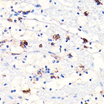
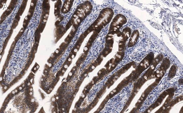
Immunohistochemistry on acetone fixed cryostat sections: 0.5-10 μg/mL using an APAAP or PAP procedure. Antibody also reacts with 4% PFA fixed frozen tissue. Typical block contains 1% non-fat dry milk or 3% NGS and 0.2-1.5% triton X-100 {higher triton is only used on thick sections; blocked for 1-3 hours at room temperature. Primary antibody is diluted only in PBS or TBS without excess blocking materials (no triton or milk); typical dilution is 1:200-1:1000 with incubations up to 48 hours at 4C. Washed 3x10′ in PBS and detect via DAB. Immunoreactive neurons are characterized by the dark brown, fine granular deposition in soma and proximal dendrites.

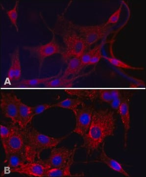
Immunocytochemistry on cell smears: 0.5-10 μg/mL.

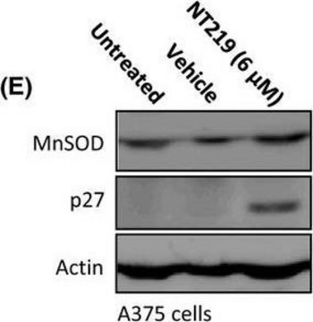
Western blot: MnS1 antibody will recognize at 25kDa band under reducing conditions; this is the monomer of the MnSOD protein.

Optimal working dilutions must be determined by end user.
Research Category
Neuroscience
Research Sub Category
Oxidative Stress
This Anti-MnSOD Antibody, clone MnS-1 is validated for use in WB, IC, IH for the detection of MnSOD.

Physical form

Format: Purified
Purified immunoglobulin. Liquid in PBS.

Storage and Stability

Prior to opening maintain at 2-8°C for up to six month after date of receipt. After opening maintain at -20°C in undiluted aliquots for up to six months. Avoid repeated freeze/thaw cycles.

Other Notes

Concentration: Please refer to the Certificate of Analysis for the lot-specific concentration.

Legal Information

CHEMICON is a registered trademark of Merck KGaA, Darmstadt, Germany

Disclaimer

Unless otherwise stated in our catalog or other company documentation accompanying the product(s), our products are intended for research use only and are not to be used for any other purpose, which includes but is not limited to, unauthorized commercial uses, in vitro diagnostic uses, ex vivo or in vivo therapeutic uses or any type of consumption or application to humans or animals.

Not finding the right product?  

Try our Product Selector Tool.

Storage Class

12 - Non Combustible Liquids

wgk_germany

WGK 2

flash_point_f

Not applicable

flash_point_c

Not applicable


Certificates of Analysis (COA)

Search for Certificates of Analysis (COA) by entering the products Lot/Batch Number. Lot and Batch Numbers can be found on a product’s label following the words ‘Lot’ or ‘Batch’.

Already Own This Product?

Find documentation for the products that you have recently purchased in the Document Library.

Visit the Document Library

Aleksandra Jankovic et al.
Free radical research, 50(sup1), S51-S63 (2016-09-07)
Setting the correct ratio of superoxide anion (O2•-) and nitric oxide (•NO) radicals seems to be crucial in restoring disrupted redox signaling in diabetic skin and improvement of •NO physiological action for prevention and treatment of skin injuries in diabetes.
Proteomic analysis of radiation-induced changes in rat lung: Modulation by the superoxide dismutase mimetic MnTE-2-PyP(5+).
Yakovlev, VA; Rabender, CS; Sankala, H; Gauter-Fleckenstein, B; Fleckenstein et al.
International Journal of Radiation Oncology, Biology, Physics null
Emily E Brown et al.
Redox biology, 24, 101201-101201 (2019-05-01)
Age-related macular degeneration (AMD) is the leading cause of vision loss in the western world. Recent evidence suggests that RPE and photoreceptors have an interconnected metabolism and that mitochondrial damage in RPE is a trigger for degeneration in both RPE
Marie Bergeaud et al.
Cell cycle (Georgetown, Tex.), 12(17), 2781-2793 (2013-08-24)
We and others previously reported that endogenous p53 can be located at mitochondria in the absence of stress, suggesting that p53 has a role in the normal physiology of this organelle. The aim of this study was to characterize in
Xiaoxing Feng et al.
Molecular cancer, 11, 48-48 (2012-07-31)
Cell death induced by poly(ADP-ribose) (PAR) and mediated by apoptosis-inducing factor (AIF) is well-characterized in models of ischemic tissue injury, but their roles in cancer cell death after chemotherapy are less understood. Here we investigated the roles of PAR and

Our team of scientists has experience in all areas of research including Life Science, Material Science, Chemical Synthesis, Chromatography, Analytical and many others.

Contact Technical Service