Skip to Content
MilliporeSigma
All Photos(1)

Key Documents

305R-1

Sigma-Aldrich

Cytokeratin 5 (EP1601Y) Rabbit Monoclonal Primary Antibody

Sign Into View Organizational & Contract Pricing

Select a Size


Select a Size

Change View

About This Item

UNSPSC Code:
12352203
NACRES:
NA.41

biological source

rabbit

Quality Level

100
500

conjugate

unconjugated

antibody form

culture supernatant

antibody product type

primary antibodies

clone

EP1601Y, monoclonal

description

For In Vitro Diagnostic Use in Select Regions (See Chart)

form

buffered aqueous solution

species reactivity

human

packaging

vial of 0.1 mL concentrate (305R-14)
vial of 0.5 mL concentrate (305R-15)
bottle of 1.0 mL predilute (305R-17)
vial of 1.0 mL concentrate (305R-16)
bottle of 7.0 mL predilute (305R-18)

General description

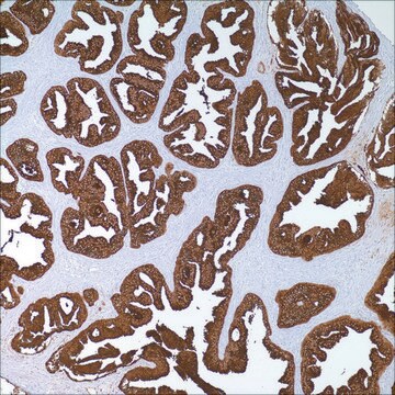
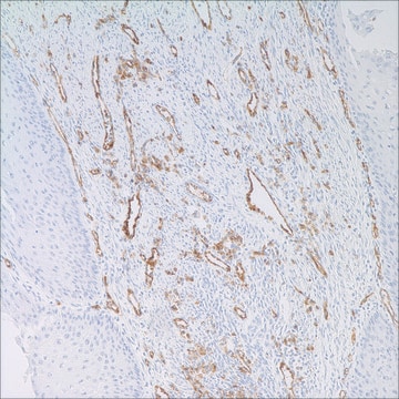
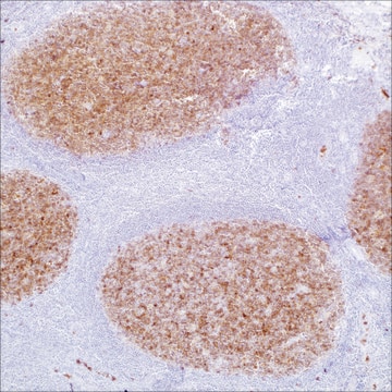
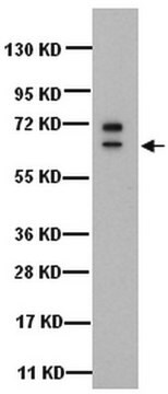
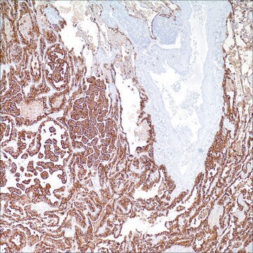
Cytokeratin 5 is an intermediate filament protein of 58 kD molecular weight within the cytokeratin family. It is a type II (basic) cytokeratin. Antibodies to this protein identify basal cells of squamous and glandular epithelia, myoepithelia, and mesothelium. Anti-cytokeratin 5 has been useful in the differential diagnosis of metastatic carcinoma in the pleura versus epithelioid mesothelioma. Epithelioid mesotheliomas are strongly positive in almost all cases, but up to 11% of pulmonary adenocarinomas will show focal immunoreactivity. Almost all squamous cell carcinomas, half of transitional carcinomas, and many undifferentiated large cell carcinomas immunostain with anti-CK 5. Anti-CK 5, along with anti-p63, affords a high sensitivity and specificity for squamous differentiation. Myoepithelial cells of the breast, glandular epithelia, and basal cells of the prostate are labeled with anti-CK 5. This antibody, along with anti-CK 14, has found application in identifying basal-like breast carcinoma, a tumor with poor prognosis. Some carcinomas of ovarian origin may display anti-CK 5 positivity.

Quality


IVD

IVD

IVD

RUO

Linkage

Cytokeratin 5 Positive Control Slides, Product No. 305S, are available for immunohistochemistry (formalin-fixed, paraffin-embedded sections).

Physical form

Solution in Tris Buffer, pH 7.3-7.7, with 1% BSA and <0.1% Sodium Azide

Preparation Note

Download the IFU specific to your product lot and formatNote: This requires a keycode which can be found on your packaging or product label.

Other Notes

For Technical Service please contact: 800-665-7284 or email: service@cellmarque.com

Legal Information

Cell Marque is a trademark of Merck KGaA, Darmstadt, Germany

Not finding the right product?  

Try our Product Selector Tool.

Storage Class

12 - Non Combustible Liquids

wgk_germany

WGK 2

flash_point_f

Not applicable

flash_point_c

Not applicable


  • Choose from one of the most recent versions:

    Certificates of Analysis (COA)

    Lot/Batch Number

    Don't see the Right Version?

    If you require a particular version, you can look up a specific certificate by the Lot or Batch number.

    Already Own This Product?

    Find documentation for the products that you have recently purchased in the Document Library.

    Visit the Document Library

    A Douglas-Jones et al.
    Histopathology, 47(2), 202-208 (2005-07-28)
    To investigate agreement on core biopsy diagnosis of papillary breast lesions, which is acknowledged as a difficult area, and to determine the effect of the use of immunohistochemistry (IHC) to assist diagnosis. Haematoxylin and eosin (H&E) sections of 129 core
    David J Dabbs et al.
    Modern pathology : an official journal of the United States and Canadian Academy of Pathology, Inc, 19(11), 1506-1511 (2006-08-31)
    The basal phenotype of breast carcinoma was demonstrated from a study of gene expression profiles, which demonstrated five carcinoma phenotypes with differing immunohistologic profiles and outcomes. The basal phenotype, so-named because of an immunohistologic profile that is similar to myoepithelial
    Catherine L Clarke et al.
    The Journal of pathology, 204(2), 147-152 (2004-09-18)
    In recent studies, Böcker and colleagues described a population of cells in paraffin wax sections of normal human breast that express cytokeratins (CK) 5/6 without expression of CK8/18 or smooth muscle actin (SMA). They proposed that these represent stem cells
    Camilla E Comin et al.
    The American journal of surgical pathology, 31(8), 1139-1148 (2007-08-02)
    Distinguishing between epithelioid peritoneal mesothelioma and papillary serous carcinomas involving the peritoneum may be very difficult, owing to overlapping morphologic features. Immunohistochemistry may facilitate establishing a correct diagnosis, but, as no single antibody has demonstrated absolute sensitivity and specificity for
    Chad A Livasy et al.
    Human pathology, 38(2), 197-204 (2007-01-20)
    Microarray profiling of invasive breast carcinomas has identified subtypes including luminal A, luminal B, HER2-overexpressing, and basal-like. The poor-prognosis, basal-like tumors have been immunohistochemically characterized as estrogen receptor (ER)-negative, HER2/neu-negative, and cytokeratin 5/6-positive and/or epidermal growth factor receptor (EGFR)-positive. The

    Our team of scientists has experience in all areas of research including Life Science, Material Science, Chemical Synthesis, Chromatography, Analytical and many others.

    Contact Technical Service