M5284
IgG1 Isotype Control from murine myeloma
clone MOPC 21, purified immunoglobulin, buffered aqueous solution
Synonym(s):
Isotype control antibody
Sign Into View Organizational & Contract Pricing
All Photos(1)
About This Item
UNSPSC Code:
12352203
NACRES:
NA.46
Recommended Products
biological source
mouse
Quality Level
conjugate
unconjugated
antibody form
purified immunoglobulin
clone
MOPC 21, monoclonal
form
buffered aqueous solution
technique(s)
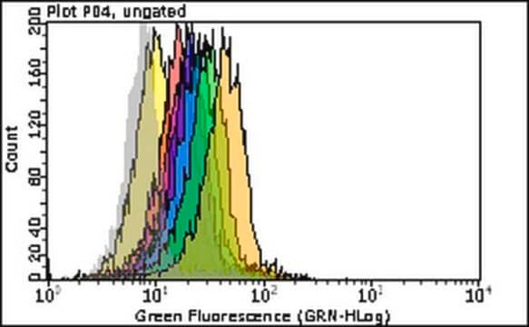
flow cytometry: suitable
shipped in
wet ice
storage temp.
2-8°C
target post-translational modification
unmodified
Looking for similar products? Visit Product Comparison Guide
General description
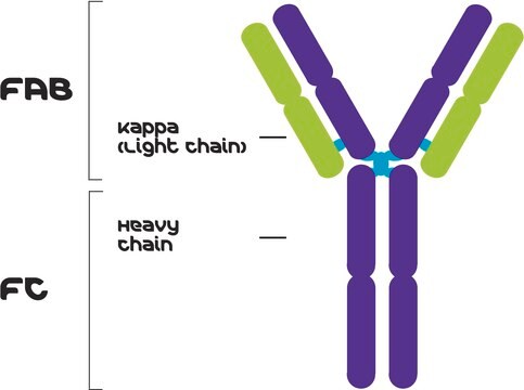
Immunoglobulin G (IgG) belongs to the immunoglobulin family and is a widely expressed serum antibody. An immunoglobulin has two heavy chains and two light chains connected by a disulfide bond. It is a glycoprotein. IgG is a major class of immunoglobulin. Mouse consists of five immunoglobulin classes- IgM, IgG, IgA, IgD and IgE. Mouse IgG is further divided into five classes- IgG1, IgG2a, IgG2b and IgG3.
Specificity
IgG1 isotype control antibody is specific for anti-mouse whole serum and anti-mouse IgG1. The product does not react with antisera to mouse IgA, IgM, IgG2a, IgG2b, and IgG3.
Application
IgG1 Isotype Control from murine myeloma has been used in:
extracellular DNA fluorescence assay
extracellular DNA fluorescence assay
- reactive oxygen species (ROS) production analysis
- immunohistochemistry
- double labelling immunohistochemistry
- immunofluorescence
- flow cytometry
- immunoprecipitation
IgG1 isotype control antibodies are suitable for use in immunohistochemistry (7μg/ml) and immunoblotting. The product may also be used for flow cytometry.
Biochem/physiol Actions
Immunoglobulin G (IgG) is a glycoprotein antibody that regulates immune responses such as phagocytosis and is also involved in the development of autoimmune diseases . Mouse IgGs have four distinct isotypes, namely, IgG1, IgG2a, IgG2b, and IgG3. IgG1 regulates complement fixation in mice . I
Immunoglobulin G (IgG) participates in hypersensitivity type II and type III reactions. It mainly helps in immune defense. IgG helps in opsonization, complement fixation and antibody dependent cell mediated cytotoxicity.
Physical form
Solution in 0.01 M phosphate buffered saline, pH 7.4, containing 1% bovine serum albumin and 15 mM sodium azide
Disclaimer
Unless otherwise stated in our catalog or other company documentation accompanying the product(s), our products are intended for research use only and are not to be used for any other purpose, which includes but is not limited to, unauthorized commercial uses, in vitro diagnostic uses, ex vivo or in vivo therapeutic uses or any type of consumption or application to humans or animals.
Storage Class
10 - Combustible liquids
wgk_germany
WGK 3
flash_point_f
Not applicable
flash_point_c
Not applicable
Choose from one of the most recent versions:
Certificates of Analysis (COA)
Lot/Batch Number
Don't see the Right Version?
If you require a particular version, you can look up a specific certificate by the Lot or Batch number.
Already Own This Product?
Find documentation for the products that you have recently purchased in the Document Library.
Customers Also Viewed
Cathepsins B, L and D in inflammatory bowel disease macrophages and potential therapeutic effects of cathepsin inhibition in vivo
Menzel K, et al.
Clinical Immunology (Orlando, Fla.), 146(1), 169-180 (2006)
The Laboratory Rat (1998)
Molecular Genetics of Immunoglobulin, 17 (1987)
Megan S Inkeles et al.
JCI insight, 1(15), e88843-e88843 (2016-10-05)
Transcriptome profiles derived from the site of human disease have led to the identification of genes that contribute to pathogenesis, yet the complex mixture of cell types in these lesions has been an obstacle for defining specific mechanisms. Leprosy provides
Lingyun Chen et al.
Journal of agricultural and food chemistry, 54(15), 5577-5582 (2006-07-20)
Parvalbumin is a calcium-binding muscle protein that is highly conserved across fish species and amphibians. It is the major cross-reactive allergen associated with both fish and frog allergy. We used two-dimensional electrophoretic and immunoblotting techniques to investigate the utility of
Our team of scientists has experience in all areas of research including Life Science, Material Science, Chemical Synthesis, Chromatography, Analytical and many others.
Contact Technical Service